You navigate the world with a constant, silent stream of information, a breathtaking symphony of light and shadow, color and form. It’s so effortless, so innate, that you rarely stop to consider the sheer marvel that is your own vision. Yet, behind that seemingly simple act of “seeing” lies a complex biological masterpiece, a testament to millions of years of evolution. You are about to embark on a journey to unravel the wonders of human vision, to understand the intricate dance of light, optics, and neural processing that paints your reality.
Your eye, that small, seemingly unassuming orb nestled in your skull, is a sophisticated camera. Its architecture is a marvel of biological engineering, designed to capture and process light with incredible precision. Understanding its components is the first step in appreciating the magic of sight.
The Protective Outer Layer: Cornea and Sclera
Your eye’s first line of defense is its robust outer shell. The cornea, a transparent, dome-shaped structure at the front of your eye, acts like a windowpane. It’s not just protective; it’s also your eye’s primary refractive surface, bending incoming light rays even before they enter. This bending is crucial for focusing the image onto your retina. Beneath the cornea, the sclera, the familiar white of your eye, provides structural integrity and shields the delicate inner workings from damage. Think of it as the sturdy casing of your personal camera.
The Aperture Control: Iris and Pupil
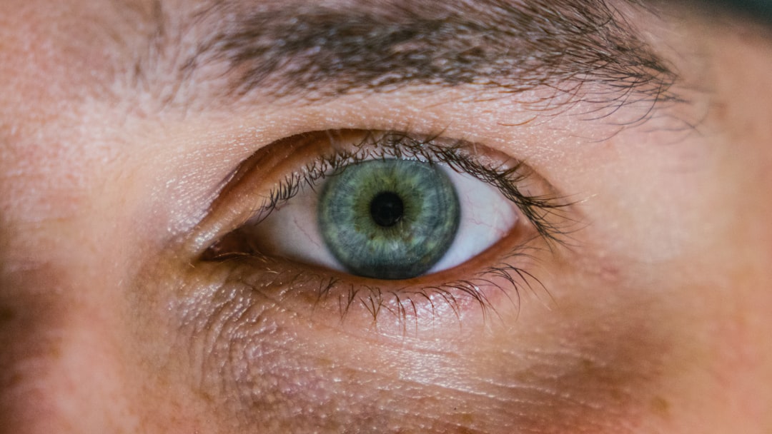
Just behind the cornea lies the iris, the colorful part of your eye – the blue, green, brown, or hazel that gives you your unique look. The iris isn’t just for aesthetics; it’s a muscular diaphragm that controls the size of the pupil, the black opening in its center. You can observe this in action: in bright light, your pupils constrict, becoming tiny pinpricks to limit the amount of light entering. In dim conditions, they dilate, widening like a camera’s aperture to capture every available photon. This dynamic regulation ensures you have optimal vision across a vast range of lighting conditions, preventing overexposure in bright sun and maximizing light gathering in the dark.
The Focusing Lens: The Crystalline Lens
Following the pupil, you encounter the crystalline lens. This transparent, flexible structure is the second major refractive component of your eye, working in tandem with the cornea. However, unlike the fixed curvature of the cornea, the lens can actively change its shape through a process called accommodation. Tiny muscles called ciliary muscles surround the lens. When you look at distant objects, these muscles relax, and the lens flattens. When you shift your gaze to something nearby, the ciliary muscles contract, causing the lens to bulge and become more convex. This remarkable adaptability allows your eye to focus light from objects at varying distances with remarkable sharpness. This is analogous to how a photographer adjusts the focus ring on their camera.
The Inner Chamber: Aqueous and Vitreous Humor
The space within your eye is filled with fluid. The aqueous humor occupies the anterior chamber, between the cornea and the iris, and the posterior chamber, between the iris and the lens. This fluid nourishes the cornea and lens, which lack their own blood supply. Behind the lens, the vast majority of your eyeball is filled with the vitreous humor, a gel-like substance that helps maintain the eye’s spherical shape and keeps the retina pressed against the back wall.
For a deeper understanding of the complexities of human vision, you may find the article on the science of perception particularly enlightening. It explores how our brains interpret visual information and the fascinating processes involved in seeing the world around us. To read more about this topic, visit Freaky Science.
The Light Catcher: The Retina’s Marvelous Workings
Once light has been focused by the cornea and lens, it travels through the vitreous humor to reach the retina, the light-sensitive tissue lining the back of your eye. This is where the true magic of converting light into electrical signals begins.
The Photoreceptor Cells: Rods and Cones
The retina is densely packed with millions of specialized cells called photoreceptors. You have two main types: rods and cones.
Rods: The Masters of Low Light
Your rods are incredibly sensitive to light, excelling in dim conditions. They are responsible for your peripheral vision and your ability to see in black and white, or more accurately, shades of gray. While they don’t detect color, their sheer number (around 120 million per eye) makes them essential for navigating in twilight and at night. They contain a pigment called rhodopsin, which undergoes a chemical change when exposed to light, triggering a signal.
Cones: The Color Detectors and Detail Sharpeners
Your cones, on the other hand, require more light to function but are responsible for your sharpest vision and your perception of color. You have three types of cones, each sensitive to different wavelengths of light: red, green, and blue. By combining the signals from these three types of cones, your brain can perceive the entire spectrum of colors. The highest concentration of cones is found in the fovea, a small depression in the center of the retina, which is your area of highest visual acuity – the part you use when you need to see fine details.
The Inner Workings: Bipolar Cells, Ganglion Cells, and the Optic Nerve
The signals generated by the rods and cones don’t travel directly to your brain. They pass through a network of other neurons within the retina. Bipolar cells receive signals from the photoreceptors and transmit them to ganglion cells. These ganglion cells are the final output neurons of the retina. Their long, slender axons bundle together to form the optic nerve, a vital cable that carries the visual information from your eye to your brain.
The Blind Spot: A Curious Omission
As the optic nerve leaves your eye, it creates a small area on the retina that lacks photoreceptor cells. This is known as the optic disc, or more commonly, the blind spot. You don’t typically notice this blind spot because your brain intelligently fills in the missing information using data from the surrounding areas and the input from your other eye. It’s a fascinating example of how your brain actively constructs your perception of reality.
The Journey to the Brain: Neural Pathways and Processing

The optic nerve is not the end of your visual journey; it’s merely the beginning of a complex neural highway. The electrical impulses generated in your retina are transmitted to your brain for interpretation.
The Thalamus: The Visual Relay Station
The optic nerves from both eyes meet at a crucial junction called the optic chiasm, located at the base of your brain. Here, the fibers from the inner half of each retina cross over, meaning that the left side of your brain receives visual information from the right half of both your visual fields, and vice versa. After the chiasm, the majority of the visual information travels to the lateral geniculate nucleus (LGN), a part of the thalamus. The LGN acts as a sophisticated relay station, sorting and organizing the visual input before sending it to the visual cortex.
The Visual Cortex: Where Sight Comes Alive
The primary destination for visual information is the primary visual cortex (V1), located in the occipital lobe at the back of your brain. This area is organized in a retinotopic map, meaning that adjacent areas in V1 process information from adjacent areas on your retina. From V1, the visual information is further processed in a hierarchical manner through a series of specialized visual areas (V2, V3, V4, V5, etc.) that are responsible for analyzing different aspects of the visual scene, such as shape, color, motion, and depth.
The Dorsal and Ventral Streams: “Where” and “What” Pathways
Two major pathways emerge from the primary visual cortex, allowing your brain to process different aspects of what you see:
The Dorsal Stream: The “Where” Pathway
This pathway, also known as the magnocellular pathway, extends upwards towards the parietal lobe. It’s primarily concerned with spatial information, motion, and the location of objects in space. It helps you understand where things are relative to you and how they are moving. This pathway is crucial for actions like reaching for an object or navigating your environment.
The Ventral Stream: The “What” Pathway
This pathway, also known as the parvocellular pathway, extends downwards towards the temporal lobe. It’s responsible for identifying objects, recognizing faces, and processing color information. It allows you to distinguish a cat from a dog, recognize a friend’s face, or appreciate the vibrant hues of a sunset.
The Illusion of Reality: Perception and Interpretation

Your brain doesn’t just passively receive visual data; it actively interprets and constructs your perception of reality. This is where the true wonder of vision lies – in the way your brain makes sense of the world.
Filling in the Gaps: The Brain’s Creative Power
As mentioned with the blind spot, your brain is remarkably adept at filling in missing information. It uses context, memory, and predictions to create a coherent visual experience. Optical illusions are a perfect illustration of this. They exploit the brain’s assumptions and shortcuts, demonstrating that what you see is not always a direct representation of reality but rather your brain’s best guess.
Recognizing Patterns and Objects: The Power of Prior Experience
Your ability to instantly recognize a familiar face, a favorite book, or a well-trodden path is a testament to the power of pattern recognition. Your brain has built a vast library of visual memories and uses them to categorize and understand new visual input. This is why a child learning to identify animals requires repeated exposure and consistent labeling.
The Subjectivity of Sight: Color and Emotion
Color is not an inherent property of objects; it’s a perception created by your brain in response to different wavelengths of light. Furthermore, your emotional state and past experiences can influence how you perceive colors and even entire scenes. A place you associate with happy memories might appear more vibrant and inviting, while a place linked to negative experiences might seem duller or more menacing.
Depth Perception: Judging Distances
Your brain uses a variety of cues to perceive depth and distance, allowing you to navigate your three-dimensional world. These cues include:
Binocular Cues: The Power of Two Eyes
- Retinal Disparity: Because your eyes are positioned a few inches apart, they receive slightly different images of the world. Your brain compares these two images and uses the difference (disparity) to calculate distance. This is why holding your finger up and closing one eye, then the other, makes your finger appear to jump.
- Convergence: The degree to which your eyes turn inward to focus on an object also provides depth information. The more your eyes converge, the closer the object.
Monocular Cues: Even with One Eye
- Linear Perspective: Parallel lines, such as railway tracks, appear to converge in the distance.
- Relative Size: If you know the typical size of an object (like a person), you can infer its distance based on how small it appears.
- Interposition (Overlap): If one object partially blocks the view of another, the occluding object is perceived as being closer.
- Texture Gradient: Textures appear finer and less detailed as they recede into the distance.
- Motion Parallax: As you move, closer objects appear to move faster and in the opposite direction of your movement, while distant objects appear to move slower or not at all.
Human vision is a fascinating topic that encompasses the intricate processes by which our eyes and brain work together to perceive the world around us. For those interested in delving deeper into this subject, a related article can be found at Freaky Science, which explores the complexities of visual perception and how various factors influence what we see. Understanding these mechanisms not only enhances our appreciation of sight but also sheds light on the challenges faced by those with visual impairments.
The Future of Vision: Innovations and Possibilities
| Aspect | Details |
|---|---|
| Visual Acuity | The sharpness of vision, measured by the ability to discern letters or numbers at a given distance. |
| Field of View | The extent of the observable world that is seen at any given moment without moving the eyes. |
| Color Vision | The ability to perceive different colors, typically enabled by cone cells in the retina. |
| Depth Perception | The ability to perceive the relative distance of objects in the visual field. |
| Visual Processing | The complex neurological process by which the brain interprets visual information from the eyes. |
The journey into understanding human vision is far from over. Scientists and engineers are continuously pushing the boundaries, leading to exciting innovations that promise to enhance, restore, and even augment our sight.
Restoring Sight: Advances in Ophthalmology
The field of ophthalmology has made incredible strides in treating conditions that impair vision. From advanced surgical techniques for cataracts and refractive errors (like LASIK) to gene therapies for inherited retinal diseases and prosthetic retinas, the aim is to restore sight to those who have lost it. These technologies offer a beacon of hope and are continually improving.
Augmenting Vision: Beyond Natural Capabilities
Beyond restoration, there’s a growing interest in visual augmentation. Imagine contact lenses that can display information directly into your field of vision, or glasses that can enhance your ability to see in low light or filter out specific visual distractions. We’re already seeing early examples of this with smart glasses and augmented reality (AR) devices, hinting at a future where your vision can be extended and enhanced in ways we can only begin to imagine.
Understanding and Treating Neurological Visual Disorders
Research into the brain’s visual processing is also leading to better understanding and treatment of neurological disorders that affect sight, such as amblyopia (lazy eye), visual agnosias, and even the visual disturbances associated with conditions like stroke or Alzheimer’s disease. By unraveling the complex neural pathways, we can develop more targeted and effective interventions.
Your ability to see is a profound gift, a constant interplay of biology, physics, and neuroscience. From the light entering your eye to the complex interpretations happening in your brain, it’s a process so intricate and elegant that it truly is one of nature’s greatest wonders. As you go about your day, take a moment to appreciate this silent, extraordinary symphony that paints your world, allowing you to connect, explore, and experience the vastness of existence. You are not just looking at the world; you are actively creating your perception of it, a truly remarkable feat of human engineering.

Why Your Brain Had to Invent Magenta
FAQs
What is human vision?
Human vision is the ability of the eyes and brain to process visual information from the surrounding environment. It involves the perception of light, color, depth, and motion.
How does human vision work?
Human vision works through the process of light entering the eye and being focused by the cornea and lens onto the retina. The retina contains photoreceptor cells called rods and cones, which convert the light into electrical signals that are sent to the brain via the optic nerve.
What are the different parts of the human eye involved in vision?
The human eye consists of several parts that play a role in vision, including the cornea, lens, iris, retina, and optic nerve. Each part has a specific function in the process of capturing and transmitting visual information to the brain.
What are the common vision problems that people experience?
Common vision problems include nearsightedness (myopia), farsightedness (hyperopia), astigmatism, and age-related conditions such as presbyopia and cataracts. These conditions can affect the ability to see clearly at various distances.
How can people maintain good vision health?
Maintaining good vision health involves regular eye exams, wearing appropriate eyewear if needed, protecting the eyes from UV radiation, maintaining a healthy diet rich in nutrients that support eye health, and avoiding habits that can strain the eyes, such as excessive screen time.