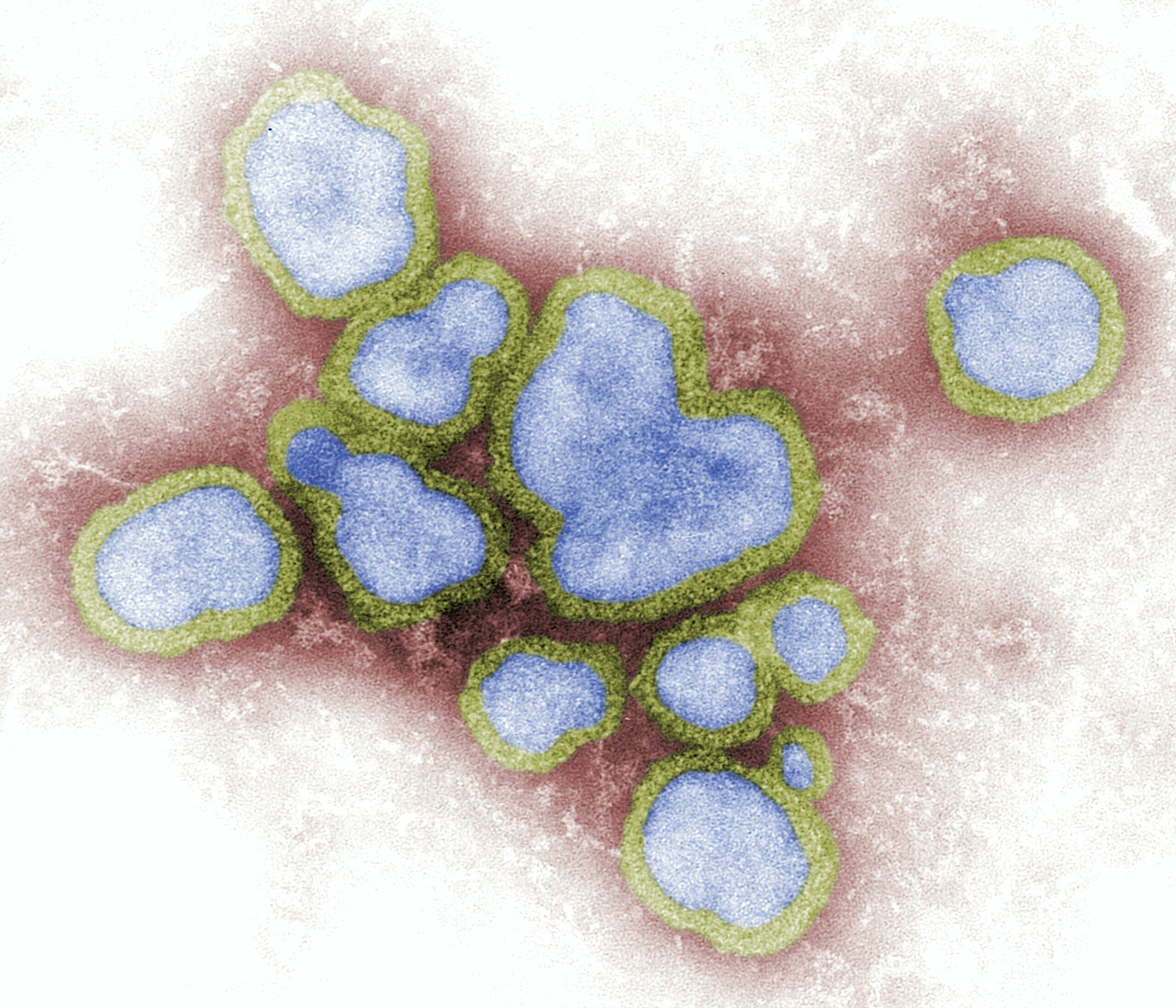
Cerebrospinal fluid (CSF) biomarkers hold significant promise in the early and accurate diagnosis of prion diseases, a group of rare and fatal neurodegenerative disorders. These biomarkers, essentially molecular messengers within the CSF, can reflect the underlying pathological processes occurring in the brain, offering a window into the disease’s progression before overt clinical symptoms become apparent. Prion diseases, characterized by the misfolding and aggregation of the prion protein (PrP), lead to neuronal dysfunction and death. Traditionally, definitive diagnosis relied on post-mortem examination of brain tissue. However, the development and refinement of CSF biomarkers are revolutionizing diagnostic capabilities, paving the way for more timely interventions, improved patient management, and enhanced understanding of these devastating conditions.
The Nature of Prion Proteins
Prion diseases, also known as transmissible spongiform encephalopathies (TSEs), are a unique class of neurological disorders. The causative agents are not viruses or bacteria, but rather misfolded proteins called prions. The normal cellular prion protein, PrPC, is a well-conserved glycoprotein found abundantly in the nervous system. Its precise physiological function is still an active area of research, but it is believed to be involved in neuronal development, synaptic function, and copper metabolism. However, under certain circumstances, PrPC can undergo a conformational change into an abnormal, infectious isoform, PrPSc (scrapie PrP). This misfolded PrPSc is resistant to degradation and acts as a template, inducing normally folded PrPC molecules to adopt the aberrant conformation. This autocatalytic process leads to the accumulation of PrPSc aggregates in the brain, forming characteristic amyloid plaques and leading to spongiform degeneration – the appearance of small, empty holes in brain tissue under microscopic examination.
The Spectrum of Prion Diseases
Prion diseases manifest in various forms, each with distinct clinical presentations and etiologies. The most common human prion disease is Creutzfeldt-Jakob disease (CJD), which occurs sporadically in approximately 85% of cases. Familial forms of CJD, as well as fatal familial insomnia (FFI) and Gerstmann-Sträussler-Scheinker syndrome (GSS), are caused by inherited mutations in the PRNP gene, which encodes the prion protein. Iatrogenic CJD can arise from medical procedures, such as contaminated surgical instruments or organ transplants. Variant CJD (vCJD), now considered to have largely been eradicated due to strict controls on the food supply chain, was linked to the consumption of beef contaminated with the bovine spongiform encephalopathy (BSE) agent. While rare, these diseases share the common pathological hallmark of prion protein misfolding and aggregation, albeit with variations in the specific strain of prion and the affected brain regions.
The Diagnostic Void: Historical Limitations
Historically, the diagnosis of prion diseases has been a formidable challenge. Clinical symptoms, which can include rapid cognitive decline, motor disturbances, and psychiatric manifestations, are often unspecific, especially in the early stages. This overlap with other neurodegenerative disorders such as Alzheimer’s disease, Lewy body dementia, and frontotemporal dementia, makes clinical differentiation difficult. Neuroimaging techniques like MRI can reveal characteristic abnormalities such as cortical ribboning and basal ganglia hyperintensities, but these findings are not always present in the early disease course and can also be seen in other conditions. Electroencephalography (EEG) may show periodic sharp wave complexes, a hallmark of sporadic CJD, but this pattern is not consistently observed throughout the disease progression and has limited sensitivity. Definitive diagnosis traditionally required a brain biopsy, an invasive procedure with associated risks, or post-mortem neuropathological examination, which obviously provides no benefit to the living patient. This diagnostic lacuna meant that patients often faced a prolonged period of uncertainty and missed opportunities for symptomatic management or participation in clinical trials.
Recent advancements in the study of cerebrospinal fluid (CSF) biomarkers have significantly enhanced our understanding of prion diseases, which are neurodegenerative disorders caused by misfolded proteins. A related article that delves deeper into the implications of these biomarkers in diagnosing and monitoring prion diseases can be found at Freaky Science. This resource provides valuable insights into the latest research findings and their potential impact on clinical practices surrounding prion-related conditions.
The Lumbar Puncture: A Gateway to the Brain’s Secrets
The Role of Cerebrospinal Fluid
The cerebrospinal fluid (CSF), a clear, colorless fluid that bathes the brain and spinal cord, acts as a protective cushion and plays a vital role in nutrient supply and waste removal within the central nervous system (CNS). Crucially, CSF is in direct contact with neural tissue, making it an ideal medium for reflecting the biochemical events occurring within the brain. Changes in the composition of CSF, including the presence or concentration of specific proteins and other molecules, can serve as indicators of pathological processes. The lumbar puncture, a minimally invasive procedure where a needle is inserted into the lower back to collect CSF, therefore represents a critical diagnostic tool for neurological disorders. Obtaining CSF allows clinicians to directly sample the environment surrounding the brain, providing a more intimate view of neural health than can be achieved through blood tests alone.
CSF as a Reflective Mirror
Think of the CSF as a mirror that reflects the state of the brain. When pathological processes, such as the misfolding and aggregation of prion proteins, occur in the brain, cellular damage and altered metabolic activity lead to the release of specific molecules into the extracellular space. These molecules then diffuse into the CSF, where they can be detected and measured. The concentration and type of these released molecules can provide valuable clues about the nature and extent of the underlying pathology. This principle is fundamental to the development of CSF biomarkers for prion diseases.
The Lumbar Puncture Procedure and Safety
The lumbar puncture, also known as a spinal tap, is a routine medical procedure performed by trained healthcare professionals. It typically involves cleaning the skin over the lower spine, applying a local anesthetic, and then carefully inserting a specialized needle into the subarachnoid space. The risks associated with a lumbar puncture are generally low and include post-procedural headache, which is usually mild and transient, and, in very rare cases, bleeding or infection. The benefits of obtaining diagnostic information that can significantly impact patient management often far outweigh these risks.
Key CSF Biomarkers in Prion Disease Detection
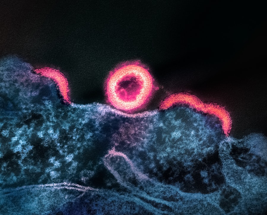
Total Tau Protein: A Marker of Neuronal Injury
Total tau (t-tau) is a highly abundant protein in the neurons of the CNS, playing a crucial role in the stabilization of microtubules, which are essential components of the neuronal cytoskeleton. Under normal conditions, tau is primarily located within neurons. However, upon neuronal damage or death, tau is released into the extracellular space and subsequently detected in the CSF. In prion diseases, the widespread neurodegeneration caused by prion accumulation leads to a significant increase in tau release, resulting in elevated t-tau levels in the CSF. This elevation is often substantial and occurs early in the disease course, making t-tau a sensitive indicator of neuronal injury. The magnitude of t-tau elevation in prion diseases is often higher than that observed in other neurodegenerative conditions like Alzheimer’s disease, providing a degree of diagnostic specificity.
Tau Isoforms and Their Significance
While total tau is a valuable marker, research is also exploring the significance of specific tau isoforms and phosphorylated tau (p-tau) in prion diseases. Different post-translational modifications of tau can arise from distinct pathological processes, and their analysis might offer further insights into the disease mechanism and progression. However, in the context of initial prion disease diagnostics, t-tau remains the most widely utilized and validated tau-based biomarker.
14-3-3 Proteins: A Rapid Indicator of Neuronal Damage
The 14-3-3 protein family comprises a group of conserved regulatory molecules found in all eukaryotic cells, with a particularly high concentration in the brain. These proteins are involved in a multitude of cellular processes, including signal transduction, cell cycle control, and apoptosis. In the context of neuronal injury, particularly acute damage, 14-3-3 proteins are rapidly released from damaged neurons into the extracellular fluid and subsequently appear in the CSF. The detection of 14-3-3 proteins in the CSF is considered a strong indicator of neuronal damage, and their levels are consistently elevated in patients with prion diseases. The 14-3-3 assay, often performed alongside tau measurements, has historically been a cornerstone of CJD diagnosis, offering a relatively rapid detection of significant neuronal insult.
The Mechanism of 14-3-3 Release
The precise mechanism by which 14-3-3 proteins are released into the CSF during prion disease is linked to the cytotoxic effects of prion aggregates and the resulting neuronal dysfunction. While the exact triggers are still being elucidated, it is understood that the pathological cascade initiated by prions leads to widespread cellular stress and compromised neuronal integrity, facilitating the release of intracellular proteins like 14-3-3.
Real-time Quaking-Induced Conversion (RT-QuIC): The Gold Standard for Prion Detection
The development of the Real-time Quaking-Induced Conversion (RT-QuIC) assay has marked a paradigm shift in prion disease diagnostics. This highly sensitive and specific technique directly detects the misfolded prion protein (PrPSc) itself, rather than just the downstream consequences of neuronal damage. RT-QuIC utilizes a process of cyclic amplification, mimicking the in vivo process of prion propagation, to detect minute amounts of PrPSc in biological samples. In essence, a small quantity of abnormal prion protein from the patient’s sample is incubated with recombinant cellular prion protein (recPrP) under specific buffer conditions that promote misfolding. Using fluorescence, the assay monitors the formation of amyloid fibrils, which are indicative of the presence of PrPSc.
The Principles of Amplification
Imagine trying to find a single, specific needle in an enormous haystack. Traditional methods might involve sifting through the entire haystack, a laborious and potentially inaccurate process. RT-QuIC, on the other hand, employs a clever amplification strategy. It’s like having a special tool that, when exposed to even a tiny fragment of the needle, can quickly create many more identical fragments. This iterative process rapidly amplifies the presence of even minuscule amounts of the target molecule, making it detectable.
RT-QuIC in CSF and Other Biofluids
While RT-QuIC has demonstrated exceptional accuracy in CSF samples, research is expanding its application to other biofluids, including nasal brushings and even blood, though sensitivity in blood remains a significant challenge. The ability to detect PrPSc directly in CSF, with extremely high sensitivity and specificity, has made RT-QuIC the current gold standard for prion disease diagnosis.
Other Potential and Emerging Biomarkers
Beyond the established biomarkers, ongoing research is exploring a multitude of other molecules in CSF that could further enhance diagnostic accuracy and provide deeper insights into disease pathogenesis.
S100B: A Glial Cell Responder
S100B is a calcium-binding protein predominantly expressed by astrocytes and other glial cells in the CNS. These cells play supportive roles for neurons, and their activation, or astrogliosis, is a common response to neuronal injury. Elevated levels of S100B in CSF indicate astrogliosis, making it a marker of general CNS damage. While less specific to prion diseases than t-tau, 14-3-3, or RT-QuIC, S100B can contribute to the overall diagnostic picture, especially when combined with other biomarkers.
GFAP: Another Marker of Astrocytic Activation
Glial fibrillary acidic protein (GFAP) is a major intermediate filament protein of astrocytes. Similar to S100B, elevated CSF GFAP levels reflect astrocytic activation and are observed in various neurological conditions, including prion diseases. Recent studies suggest that GFAP might be a particularly sensitive marker of early neuronal damage in prion diseases, potentially preceding more prominent elevations in tau.
Neurofilament Light Chain (NfL): A Universal Marker of Axonal Damage
Neurofilament light chain (NfL) is a protein component of the neuronal cytoskeleton. In cases of axonal damage, NfL is released into the extracellular space and subsequently detected in the CSF and blood. NfL is considered a pan-neurological marker of axonal injury and is elevated in a wide range of neurodegenerative and acute neurological conditions. While not specific to prion diseases, elevated NfL levels can corroborate the presence of significant neurodegeneration and its trajectory over time.
Clinical Applications and Diagnostic Pathways
The Diagnostic Algorithm: Combining Biomarkers
The diagnosis of prion diseases is not typically reliant on a single biomarker. Instead, a diagnostic algorithm is employed, where results from multiple CSF biomarkers, alongside clinical presentation and neuroimaging findings, are integrated to arrive at the most accurate diagnosis. A typical diagnostic pathway might involve:
- Clinical Suspicion: A patient presenting with rapidly progressive dementia, myoclonus, or other suggestive neurological signs triggers further investigation.
- Neuroimaging: MRI of the brain is performed to identify characteristic changes.
- Lumbar Puncture: CSF is collected for biomarker analysis.
- Biomarker Analysis: Tests for t-tau, 14-3-3 proteins, and crucially, RT-QuIC are performed.
- Integration of Findings: The results are synthesized. A characteristic clinical picture, suggestive MRI findings, and a combination of elevated t-tau, positive 14-3-3, and a highly positive RT-QuIC assay strongly support a diagnosis of prion disease.
The sensitivity and specificity of these combined biomarkers are remarkably high, allowing for accurate diagnosis in a significant proportion of suspected cases.
Differential Diagnosis: Distinguishing from Other Conditions
The diagnostic utility of CSF biomarkers extends crucially to distinguishing prion diseases from other neurological disorders that can mimic their clinical presentation. As mentioned earlier, conditions such as Alzheimer’s disease, Lewy body dementia, and autoimmune encephalitis can present with cognitive decline and other neurological symptoms. However, the specific patterns of biomarker abnormalities can help differentiate these conditions. For instance, while tau is elevated in Alzheimer’s disease, the levels are typically lower than those seen in prion diseases, and the RT-QuIC assay will be negative, as Alzheimer’s is not a prion disease. Similarly, autoimmune encephalitis, which involves an immune attack on the brain, will often have a different biomarker profile and may respond to immunosuppressive therapies, unlike prion diseases.
Resource Allocation and Public Health Implications
The ability to diagnose prion diseases accurately and in a timely manner has significant implications for public health and resource allocation. For individuals and families, a definitive diagnosis brings closure and allows for appropriate care planning and support. For healthcare systems, accurate diagnosis prevents unnecessary and costly investigations into other conditions. Furthermore, in the context of animal health and potential zoonotic transmission (though extremely rare in humans), robust diagnostic capabilities are essential for surveillance and control measures.
Recent advancements in the study of cerebrospinal fluid biomarkers for prion diseases have opened new avenues for early diagnosis and monitoring of these neurodegenerative conditions. Researchers are increasingly focusing on the role of specific proteins and their alterations in the cerebrospinal fluid, which may serve as crucial indicators of prion pathology. For further insights into this topic, you can explore a related article that discusses the implications of these biomarkers in greater detail at Freaky Science. This growing body of research not only enhances our understanding of prion diseases but also paves the way for potential therapeutic strategies.
Therapeutic Implications and Future Directions
| Biomarker | Type | Diagnostic Use | Sensitivity (%) | Specificity (%) | Notes |
|---|---|---|---|---|---|
| 14-3-3 Protein | Protein | Supportive for Creutzfeldt-Jakob Disease (CJD) | 85-95 | 80-90 | Widely used but not specific to prion diseases |
| Tau Protein (Total Tau) | Protein | Indicative of neuronal damage in prion diseases | 90-95 | 85-90 | Elevated in CJD and other neurodegenerative diseases |
| RT-QuIC (Real-Time Quaking-Induced Conversion) | Assay detecting prion seeding activity | Definitive diagnosis of prion diseases | 92-98 | 99-100 | High specificity and sensitivity; gold standard |
| S100B Protein | Protein | Marker of astrocytic activation | Variable | Variable | Less commonly used; supportive marker |
| Neuron-Specific Enolase (NSE) | Protein | Marker of neuronal injury | 70-85 | 75-85 | Elevated in prion and other neurodegenerative diseases |
The Quest for Treatments
Currently, there are no approved treatments that can cure or halt the progression of prion diseases. The rapid neurodegeneration leaves little room for intervention once significant damage has occurred. However, the development and refinement of CSF biomarkers are crucial for the success of future therapeutic strategies.
Biomarkers in Clinical Trials
As researchers strive to develop novel therapies, CSF biomarkers will play a pivotal role in clinical trials. They can serve as:
- Inclusion/Exclusion Criteria: Ensuring that trial participants have confirmed prion disease and meet specific diagnostic criteria.
- Outcome Measures: Tracking the efficacy of experimental treatments by monitoring changes in biomarker levels over time. A successful therapy would ideally lead to a reduction in neuronal damage markers like t-tau and GFAP, or even a decrease in PrPSc detected by RT-QuIC.
- Early Detection of Response: Identifying patients who are responding to treatment earlier than would be possible through clinical assessment alone.
The ability of biomarkers to capture sub-clinical changes in the disease process opens up the possibility of intervening before irreversible neuronal damage occurs, a critical goal for any effective treatment.
Precision Medicine and Personalized Diagnostics
The future of prion disease diagnostics and therapeutics likely lies in a more personalized approach. Advanced proteomic and metabolomic analyses of CSF could uncover a more comprehensive panel of biomarkers that not only diagnose prion diseases but also predict disease trajectory, identify specific prion strains, or even reveal susceptibility to certain therapeutic interventions. This move towards precision medicine, guided by fine-tuned biomarker analysis, holds the promise of tailoring treatments to the individual patient for maximum benefit.
Challenges and Opportunities in Biomarker Research
Despite the remarkable progress, challenges remain in the field of prion disease biomarkers. These include:
- Standardization: Ensuring consistent assay performance and interpretation across different laboratories globally is crucial for comparability and robustness of diagnostic data.
- Sensitivity in Early Stages: While current biomarkers are highly sensitive, further improvements in detecting very early or pre-symptomatic prion accumulation would be invaluable for preventative strategies.
- Blood-Based Biomarkers: Developing highly sensitive and specific biomarkers in blood would be a significant advancement, as it would eliminate the need for lumbar punctures, making diagnostics more accessible and patient-friendly. While RT-QuIC in blood is showing promise, it is not yet as robust as in CSF.
- Understanding Non-Prion Mimics: Continued research into the specific biomarker profiles of conditions that mimic prion diseases will further refine differential diagnostic capabilities.
The ongoing research into CSF biomarkers for prion diseases represents a dynamic and evolving field. The continuous refinement of existing assays and the discovery of novel molecular markers are steadily improving our ability to detect and understand these challenging disorders. The journey from post-mortem diagnosis to precise, early-stage detection through a simple lumbar puncture, powered by sophisticated biomarkers, is a testament to scientific ingenuity and holds significant hope for future advancements in managing and potentially treating prion diseases. The protein messengers within the CSF are no longer silent witnesses to neural decay; they are becoming active participants in unraveling the mysteries of prion pathogenesis and guiding the path towards effective diagnostics and, hopefully, treatments.
FAQs
What are cerebrospinal fluid biomarkers for prions?
Cerebrospinal fluid (CSF) biomarkers for prions are specific proteins or molecules found in the CSF that help detect the presence of prion diseases. These biomarkers assist in diagnosing conditions like Creutzfeldt-Jakob disease by indicating abnormal prion protein activity.
How are cerebrospinal fluid biomarkers used in diagnosing prion diseases?
CSF biomarkers are analyzed through lumbar puncture to detect abnormal prion proteins or related changes. Elevated levels or specific patterns of these biomarkers can confirm the presence of prion diseases, aiding early and accurate diagnosis.
Which cerebrospinal fluid biomarkers are commonly tested for prion diseases?
Common CSF biomarkers for prion diseases include 14-3-3 protein, tau protein, and real-time quaking-induced conversion (RT-QuIC) assay results. These markers help differentiate prion diseases from other neurological disorders.
Are cerebrospinal fluid biomarkers reliable for prion disease diagnosis?
While CSF biomarkers are valuable diagnostic tools, their reliability varies. Tests like RT-QuIC have high specificity and sensitivity, but results should be interpreted alongside clinical symptoms and other diagnostic methods for accurate diagnosis.
Can cerebrospinal fluid biomarkers be used to monitor prion disease progression?
Currently, CSF biomarkers are primarily used for diagnosis rather than monitoring disease progression. Research is ongoing to determine if changes in biomarker levels can reflect disease advancement or response to potential treatments.