The human eye, a marvel of biological engineering, possesses an astonishing capacity to perceive the world. While we largely operate in daylight, our visual system is equipped with mechanisms that allow us to navigate and interpret even in the dimmest light. This ability, often referred to as human night vision, is a testament to the specialized cells within the retina: rods and cones. These photoreceptor cells, each with distinct roles and sensitivities, orchestrate our vision under varying illumination levels, with rods being the unsung heroes of our nocturnal world.
At the heart of our visual perception lies the retina, a light-sensitive tissue lining the back of the eye. Within this intricate network, millions of photoreceptor cells convert light into electrical signals that are then transmitted to the brain for interpretation. Two primary types of photoreceptors dominate this landscape: cones and rods. Understanding their fundamental differences is crucial to grasping how we see in both bright and dim conditions.
The Role of Cones in Photopic Vision
Cones are primarily responsible for our vision in bright light, a state known as photopic vision. They are concentrated in the fovea, the central part of the retina, which affords us sharp, detailed vision. Humans possess three types of cones, each sensitive to different wavelengths of light: red, green, and blue. This trichromatic nature allows us to perceive a vast spectrum of colors. When light strikes a cone, it triggers a cascade of biochemical reactions that ultimately generate an electrical signal. However, cones require a relatively high level of light to be activated and are not very sensitive to low light levels. Their acuity and color discrimination are superior to rods, making them essential for tasks requiring fine detail and vibrant hues during the day.
The Sensitivity of Rods in Scotopic Vision
In stark contrast to cones, rods are exquisitely sensitive to light. They are far more numerous than cones, with estimates suggesting around 120 million rods per eye compared to 6 million cones. Rods are distributed throughout the peripheral retina, with a scarcity in the fovea. This distribution explains why our peripheral vision is better in low light, and why trying to focus directly on a dim object often makes it disappear. Rods contain a photopigment called rhodopsin, which is highly efficient at capturing even a single photon of light. This remarkable sensitivity allows us to detect faint illuminations that would be imperceptible to cones. However, rods do not distinguish between different wavelengths of light, meaning they do not contribute to color vision. Instead, they provide monochromatic vision, rendering the world in shades of gray. This trade-off—sensitivity for color and detail—is the core of their specialization for scotopic vision, or vision in dim light.
Human vision relies on two types of photoreceptor cells in the retina: rods and cones. While cones are responsible for color vision and function best in bright light, rods are highly sensitive to low light levels, making them essential for night vision. For a deeper understanding of how these cells work together to enable us to see in varying light conditions, you can read a related article on this topic at Freaky Science.
The Process of Dark Adaptation: Enhancing Rod Sensitivity
When transitioning from a brightly lit environment to darkness, our eyes undergo a remarkable process called dark adaptation. This physiological adjustment allows our visual system, particularly the rods, to become increasingly sensitive to lower light levels, thereby unlocking our night vision capabilities. This adaptation is not instantaneous; it unfolds over a period of minutes and even hours.
The Initial Burst of Adaptation
Immediately upon entering a dim environment, our cones, which were heavily bleached by the bright light, begin to regenerate their photopigments. This initial phase of dark adaptation, lasting about 5-10 minutes, sees a significant improvement in our ability to see. We can start to discern larger shapes and outlines, and our vision in the dim light becomes more functional. During this period, the cones are still contributing, but their sensitivity is still limited.
The Prolonged Ascent of Rod Sensitivity
The true unlocking of night vision occurs as our rods enter their peak performance. After the initial 5-10 minutes, the cones reach their maximum sensitivity in the dark. However, their overall sensitivity remains significantly lower than that of the rods. The crucial phase of dark adaptation for night vision is dominated by the rods. Over the next 20-30 minutes, and sometimes up to an hour or more, the rhodopsin in the rods regenerates fully. This regeneration dramatically increases the sensitivity of the rods to light. A single photon, which would have been insufficient to stimulate a cone, can now trigger a cascade of events in a rod, leading to a discernible signal. This prolonged adaptation is why it takes time for our eyes to adjust to complete darkness, and why our vision improves gradually in a dimly lit room after entering from sunlight.
The Biochemistry of Rhodopsin Regeneration
The key player in rod sensitivity is rhodopsin, a complex molecule composed of a protein called opsin and a light-sensitive pigment called retinal. When light strikes rhodopsin, retinal undergoes a conformational change, initiating a biochemical cascade that ultimately leads to an electrical signal. In bright light, rhodopsin is bleached, meaning retinal detaches from opsin. Dark adaptation involves the enzymatic process of regenerating the bound form of retinal and reattaching it to opsin, making the rhodopsin available to absorb photons once again. This intricate biochemical dance allows the rods to become hyper-sensitive in the absence of light.
The Neural Pathways of Night Vision

The electrical signals generated by rods and cones do not directly travel to the brain. Instead, they undergo processing within the retina itself through a complex network of neurons. This intricate neural circuitry plays a vital role in enhancing the signals from the rods and preparing them for transmission to the visual cortex.
The Role of Bipolar Cells
Following the stimulation of photoreceptors, the signals are passed on to bipolar cells. There are different types of bipolar cells, and their interactions with rods and cones are crucial. For instance, some bipolar cells are excited by photoreceptors, while others are inhibited. This push-pull mechanism contributes to the enhancement of contrast and the detection of edges, which is particularly important in low-light conditions where visual information is sparse.
The Convergence of Rod Signals
A significant aspect of rod signaling is the high degree of convergence. Many rods converge onto a single bipolar cell, and many bipolar cells converge onto a single ganglion cell. This convergence means that the signals from a large number of rods are summed up. While this pooling of signals can reduce spatial resolution, it dramatically increases sensitivity. The summation allows for the detection of very faint overall light levels, as the combined excitation of multiple rods can cross the threshold for activation in the downstream neurons. This is a prime example of how our visual system sacrifices detail for enhanced sensitivity in dim light.
The Influence of Horizontal and Amacrine Cells
The retinal network also includes horizontal cells and amacrine cells. Horizontal cells play a role in lateral inhibition, which helps to sharpen the contrast between adjacent areas of light and dark. Amacrine cells are involved in more complex processing, modulating the signals from bipolar cells and influencing how information is transmitted to the ganglion cells. These interneurons contribute to the sophisticated processing of visual information within the retina, refining the signals before they are sent to the brain.
The Brain’s Interpretation of Rod Signals
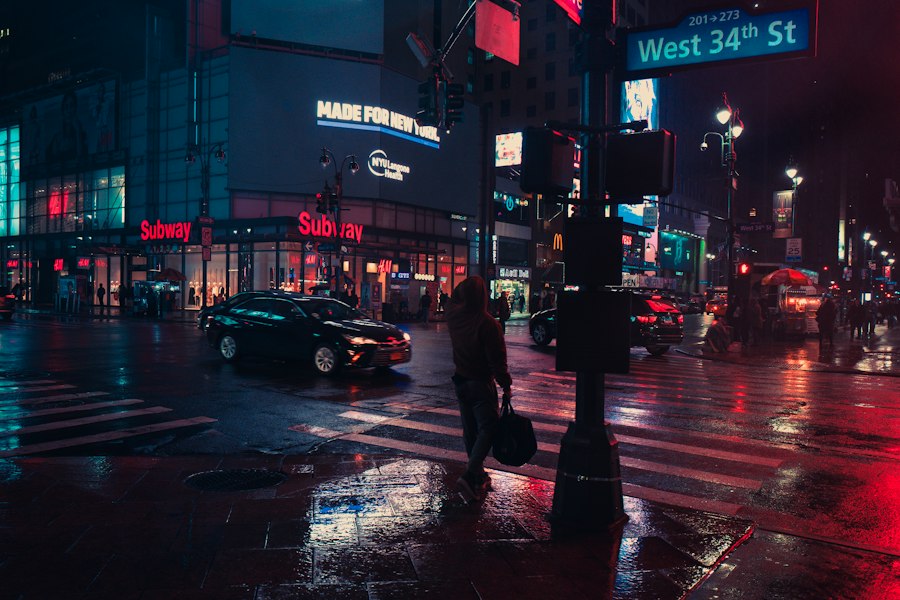
Once the processed electrical signals from the retina reach the brain via the optic nerve, they are interpreted and integrated to form our perception of the visual world. The brain’s ability to make sense of the monochromatic information provided by rods is what constitutes our experience of night vision.
The Visual Cortex and Low-Light Processing
The signals ultimately arrive at the visual cortex, the part of the brain responsible for processing visual information. While the visual cortex is capable of processing a wide range of visual input, it has specific pathways and mechanisms for interpreting the signals that originate from the rods. The brain learns to associate specific patterns of gray shades and subtle movements with objects and our surroundings.
The Absence of Color and Detail
It is important to reiterate that rod vision is monochromatic. Therefore, when we are relying on our rods for night vision, we are essentially seeing the world in shades of gray. The rich tapestry of colors we perceive during the day is absent. Furthermore, the high convergence of rod signals, while enhancing sensitivity, leads to a significant loss of fine detail and spatial resolution. Objects in dim light appear blurry and less defined compared to their appearance in bright daylight.
The Role of Perception and Expectation
Beyond the purely physiological processes, our perception of night vision is also influenced by our cognitive processes. Our brains are adept at filling in gaps and making educated guesses based on past experiences and expectations. In a familiar environment, our brain can often construct a coherent image even with limited visual input. This cognitive aspect of perception contributes to our ability to navigate and function in low-light conditions.
Human vision relies on specialized cells in the retina known as rods and cones, which play crucial roles in our ability to see in different lighting conditions. While cones are responsible for color vision and function best in bright light, rods are highly sensitive to low light levels, making them essential for night vision. For a deeper understanding of how these cells work together to enhance our visual experience, you can explore this insightful article on the topic. To learn more about the fascinating mechanisms behind human night vision, visit this article.
Factors Affecting Human Night Vision
| Category | Rods | Cones |
|---|---|---|
| Location in the retina | Peripheral retina | Central retina |
| Function | Responsible for night vision and peripheral vision | Responsible for color vision and detailed central vision |
| Sensitivity to light | High sensitivity to low light levels | Less sensitive to low light levels |
| Number in the retina | Approximately 120 million | Approximately 6 million |
While our eyes are naturally equipped for night vision, several factors can influence its effectiveness. These include physiological conditions, environmental influences, and even our lifestyle choices. Understanding these factors can help individuals optimize their natural night vision capabilities.
Age and Declining Sensitivity
As we age, there is a natural decline in both rod and cone function. This can manifest as a decreased ability to adapt to darkness and a reduced overall sensitivity to light. The regeneration of rhodopsin may slow down, and the number of photoreceptor cells might decrease. Consequently, older adults often experience more difficulty seeing in dim light.
Nutritional Deficiencies and Vitamin A
Vitamin A is a crucial component of rhodopsin. A deficiency in vitamin A can lead to a condition known as nyctalopia, or night blindness, where the ability to see in dim light is severely impaired. This underscores the importance of a balanced diet rich in vitamin A for maintaining healthy vision, particularly night vision.
Medications and Ocular Health
Certain medications and medical conditions can also impact night vision. For example, some drugs used to treat glaucoma or certain mental health conditions can affect the sensitivity of photoreceptors or interfere with neurotransmission in the retina. Eye conditions such as cataracts can scatter light and reduce the amount of light reaching the retina, thereby diminishing night vision.
Environmental Factors: Light Pollution and Visual Acuity
The presence of light pollution in urban environments can significantly impair our natural night vision. Even dim ambient light can prevent the complete regeneration of rhodopsin, making it harder for our rods to function optimally. Conversely, in truly dark environments, our rods can achieve their full potential. Furthermore, conditions that affect overall visual acuity, such as myopia (nearsightedness) or astigmatism, will also impact the clarity of vision in low light, even if the rod sensitivity is intact.
In conclusion, the remarkable ability of humans to see in dim light, our night vision, is a complex interplay of specialized photoreceptor cells, intricate neural processing, and the interpretive power of the brain. The rods, with their extraordinary sensitivity to light and reliance on rhodopsin, are the primary architects of our nocturnal visual world. The process of dark adaptation, where rhodopsin regenerates and rods become hyper-sensitive, is a testament to the biological resilience and adaptability of our visual system. While our night vision may not rival that of nocturnal animals, it is a vital and fascinating aspect of human perception, allowing us to navigate and appreciate the world even when the sun has set. Understanding the mechanisms behind this capability not only deepens our appreciation for the human eye but also highlights the importance of factors that can influence its effectiveness.

The Space Photos Are Real—But Your Eyes Would See This
FAQs
What are rods and cones in the human eye?
Rods and cones are photoreceptor cells located in the retina of the human eye. Rods are responsible for vision in low light conditions, while cones are responsible for color vision and visual acuity in bright light.
How do rods and cones contribute to night vision?
Rods are highly sensitive to light and are responsible for our ability to see in low light conditions, such as at night. They allow us to perceive shapes and movement in dimly lit environments.
What is the difference between rods and cones in terms of function?
Rods are more sensitive to light and are responsible for night vision, while cones are less sensitive to light and are responsible for color vision and visual acuity in bright light.
How do rods and cones adapt to changes in light levels?
Rods and cones adapt to changes in light levels through a process called dark adaptation. In low light conditions, the rods become more sensitive, allowing for better night vision. In bright light conditions, the cones become more active, allowing for color vision and visual acuity.
Can the function of rods and cones be affected by certain medical conditions?
Yes, certain medical conditions such as retinitis pigmentosa and age-related macular degeneration can affect the function of rods and cones, leading to vision impairment, including difficulties with night vision.