Understanding Necrosis vs. Apoptosis for Tissue Health
The maintenance of healthy tissue is a complex biological process governed by a delicate balance of cellular life and death. Within this dynamic, two primary mechanisms of cell death play critical roles: apoptosis and necrosis. While both lead to the demise of cells, their underlying causes, cellular mechanisms, and ultimate consequences for tissue health are fundamentally different. Understanding these distinctions is crucial for comprehending various physiological and pathological conditions affecting the human body.
Defining Apoptosis: The Programmed Cell Death
Apoptosis, often referred to as programmed cell death, is a highly regulated and energy-dependent process. It is an active, genetically controlled mechanism that removes unwanted, old, or damaged cells in a controlled and organized manner. This carefully orchestrated cellular suicide is essential for normal development, tissue homeostasis, and the elimination of potentially harmful cells. Apoptosis serves as a vital housekeeping function for multicellular organisms, ensuring the proper formation of tissues and organs during embryonic development, and maintaining the integrity of adult tissues throughout life.
Defining Necrosis: The Uncontrolled Cell Death
In contrast, necrosis is a form of uncontrolled cell death that occurs as a result of injury or disease. It is a passive process, typically triggered by external factors such as trauma, toxins, infection, or ischemia (lack of oxygen). Unlike apoptosis, necrosis is characterized by a loss of cellular integrity, leading to the rupture of the cell membrane and the spilling of intracellular contents into the surrounding tissue. This release of cellular debris elicits an inflammatory response, which can further damage neighboring cells and compromise tissue function.
Understanding the differences between necrosis and apoptosis is crucial for comprehending tissue health and disease progression. While necrosis often results from acute injury leading to uncontrolled cell death and inflammation, apoptosis is a regulated process that plays a vital role in maintaining cellular homeostasis. For a deeper exploration of these processes and their implications in health and disease, you can read the related article on tissue health at Freaky Science.
Triggers and Causes of Cell Death
The Diverse Inducers of Apoptosis
Apoptosis can be triggered by a multitude of internal and external stimuli. Internally, cells may initiate apoptosis in response to DNA damage that cannot be repaired, oxidative stress, or the accumulation of misfolded proteins. These signals activate specific intracellular pathways that ultimately lead to cell dismantling. Externally, signals from other cells, such as certain cytokines and hormones, can also induce apoptosis. For instance, the binding of death ligands to their corresponding receptors on the cell surface can initiate the apoptotic cascade. This programmed elimination is critical for eliminating cells that pose a threat to the organism, such as virus-infected cells or precancerous cells. Developmental processes often rely heavily on apoptosis to sculpt tissues and organs, removing unnecessary structures or shaping boundaries.
The External Insults Leading to Necrosis
Necrosis, on the other hand, is primarily a response to severe cellular injury. Physical trauma, such as blunt force or crushing injuries, can directly disrupt cell membranes and lead to necrosis. Chemical agents, including toxins and poisons, can overwhelm cellular detoxification mechanisms and cause widespread cell damage and death. Infections by bacteria, viruses, or fungi can also trigger necrosis through the release of damaging enzymes or toxins, or by eliciting a severe inflammatory response that becomes self-destructive. Perhaps one of the most common causes of necrosis in clinical settings is ischemia, a condition where a tissue is deprived of adequate blood supply and oxygen. This lack of oxygen impairs crucial cellular energy production, leading to a breakdown of cellular functions and eventual cell death.
Cellular Mechanisms and Morphology
The Intricate Pathway of Apoptosis
Apoptosis proceeds through a highly regulated cascade of molecular events. The intrinsic pathway, also known as the mitochondrial pathway, is activated by intracellular signals such as DNA damage. This leads to changes in mitochondrial membrane permeability, releasing pro-apoptotic factors like cytochrome c into the cytoplasm. Cytochrome c then activates a family of cysteine proteases called caspases, which are the executioners of apoptosis. Caspases dismantle the cell in a systematic manner, cleaving vital cellular proteins and activating enzymes that degrade DNA.
The extrinsic pathway, or death receptor pathway, is initiated by extracellular signals. Ligands like tumor necrosis factor (TNF) bind to death receptors on the cell surface, triggering a similar caspase activation cascade. Regardless of the initiating pathway, the end result is the formation of membrane-bound apoptotic bodies. These vesicles contain the
fragmented cellular components, allowing for their efficient phagocytosis by neighboring cells or specialized immune cells, such as macrophages, without causing inflammation.
The Disorganized Demise of Necrosis
Necrosis is characterized by a significant loss of cellular regulation and integrity. Unlike the controlled dismantling in apoptosis, necrotic cells swell due to the influx of water and ions, as their plasma membranes lose their barrier function. This swelling can lead to the rupture of the cell membrane, releasing the cell’s contents, including enzymes, ions, and DNA, into the extracellular space. The released intracellular components are recognized as foreign by the immune system, triggering an inflammatory response. This inflammatory process, mediated by the recruitment of leukocytes, aims to clear the cellular debris but can also cause collateral damage to surrounding healthy tissues.
Morphological Hallmarks of Necrosis and Apoptosis
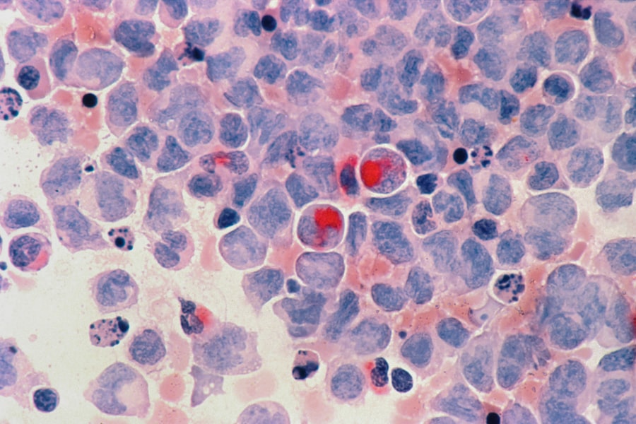
The observable changes in cellular morphology are a key distinguishing feature between necrosis and apoptosis. Apoptotic cells typically shrink, undergo chromatin condensation and nuclear fragmentation, and form distinct, membrane-bound apoptotic bodies. The cell membrane remains largely intact until the final stages of engulfment. In contrast, necrotic cells undergo swelling, the dissolution of organelles, and the loss of plasma membrane integrity, leading to widespread cellular lysis. The nucleus in necrotic cells may undergo pyknosis (condensation), karyorrhexis (fragmentation), or karyolysis (dissolution), but these changes are less organized and often accompanied by the overall disintegration of the cell.
Consequences for Tissue Health

Apoptosis: A Guardian of Tissue Homeostasis
Apoptosis plays a crucial role in maintaining tissue health and function by removing senescent, damaged, or potentially cancerous cells. During development, it sculpts organs and eliminates redundant structures. In adults, it removes cells that are no longer needed, such as the shedding of the uterine lining during menstruation, or the turnover of intestinal epithelial cells. It also eliminates cells that have sustained irreparable DNA damage, thus preventing the propagation of mutations that could lead to cancer. Furthermore, apoptosis is instrumental in immune system regulation, removing self-reactive lymphocytes that could cause autoimmune diseases.
Necrosis: A Signal of Impaired Tissue Integrity
Necrosis is invariably associated with tissue damage and dysfunction. The uncontrolled release of intracellular components triggers inflammation, which, while a necessary part of the healing process, can lead to further tissue injury if excessive or prolonged. Chronic inflammation associated with repeated necrotic events can contribute to the development of fibrotic tissue, scarring, and the loss of specialized cell function. For example, myocardial infarction (heart attack) involves extensive necrosis of heart muscle cells due to prolonged ischemia. The resulting scar tissue impairs the heart’s pumping ability. Similarly, stroke involves the necrosis of brain tissue due to lack of blood flow, leading to neurological deficits.
The Inflammatory Response: A Differentiating Factor
The presence or absence of a significant inflammatory response is a key indicator differentiating necrosis from apoptosis. Apoptosis is a “silent” form of cell death, characterized by the efficient engulfment of apoptotic bodies by phagocytes, which prevents the release of inflammatory mediators. This minimizes damage to surrounding tissues. Necrosis, on the other hand, is inherently inflammatory. The rupture of the cell membrane releases intracellular contents that act as DAMPs (damage-associated molecular patterns), activating innate immune cells and initiating a cascade of inflammatory signaling. This inflammatory response is a hallmark of necrotic tissue injury and can exacerbate tissue damage.
Understanding the differences between necrosis and apoptosis is crucial for comprehending tissue health and disease progression. While necrosis typically results from acute injury leading to cell death and inflammation, apoptosis is a programmed cell death mechanism that maintains tissue homeostasis. For a deeper exploration of these processes and their implications in various health conditions, you can refer to this insightful article on cellular mechanisms. This resource provides valuable information that can enhance your knowledge of how these two forms of cell death impact overall tissue health.
Clinical Implications and Therapeutic Significance
| Metrics | Necrosis | Apoptosis |
|---|---|---|
| Definition | Cell death due to injury or disease | Programmed cell death |
| Cell swelling | Yes | No |
| Cell fragmentation | Yes | Yes |
| Immune response | Induces inflammation | No inflammation |
| Energy requirement | No energy required | Energy required |
Apoptosis Dysregulation: Links to Disease
Dysregulation of apoptosis is implicated in a wide range of diseases. Insufficient apoptosis can contribute to the development of cancer, as cells that should be eliminated continue to proliferate. Autoimmune diseases can arise when the immune system fails to eliminate self-reactive lymphocytes through apoptosis. Conversely, excessive apoptosis can lead to the loss of vital cells, contributing to neurodegenerative diseases like Alzheimer’s and Parkinson’s, or ischemic injury. Understanding and manipulating apoptotic pathways holds significant therapeutic potential for treating these conditions.
Necrosis: Targets for Intervention
Given that necrosis signifies substantial cellular injury, therapeutic strategies often focus on preventing its occurrence or mitigating its consequences. In cases of ischemia, timely revascularization to restore blood flow is paramount to limit the extent of necrotic cell death. In conditions involving toxins or infections, interventions aim to neutralize the damaging agent and support cellular recovery. For inflammatory conditions associated with necrosis, anti-inflammatory therapies can help manage the secondary damage. Research also explores ways to promote the clearance of necrotic debris and encourage tissue regeneration.
Therapeutic Strategies Targeting Cell Death Pathways
The distinct mechanisms of apoptosis and necrosis offer different avenues for therapeutic intervention. For diseases characterized by excessive apoptosis, drugs that inhibit apoptotic pathways, such as caspase inhibitors, are being investigated. Conversely, for conditions where increased apoptosis is beneficial, such as in cancer therapy, drugs are designed to induce programmed cell death. For necrosis, interventions primarily focus on preventing the initial injury, such as thrombolytic drugs to dissolve blood clots in stroke or heart attack, or antioxidants to combat oxidative stress contributing to cell damage. Ultimately, a nuanced understanding of both apoptosis and necrosis is essential for developing effective treatments for a broad spectrum of human diseases.
FAQs
What is necrosis and apoptosis in tissue health?
Necrosis is a type of cell death that occurs as a result of injury, infection, or disease, leading to the premature death of cells and tissue damage. Apoptosis, on the other hand, is a programmed cell death that occurs as a normal part of an organism’s growth and development, as well as in response to cell damage or stress.
What are the main differences between necrosis and apoptosis?
Necrosis is characterized by swelling of the cell and organelles, followed by rupture of the cell membrane and release of cellular contents, which can lead to inflammation and damage to surrounding tissue. Apoptosis, on the other hand, is a controlled process that involves cell shrinkage, chromatin condensation, and formation of apoptotic bodies, which are then engulfed and removed by neighboring cells without causing inflammation.
How do necrosis and apoptosis impact tissue health?
Necrosis can lead to inflammation, tissue damage, and loss of function, as the release of cellular contents triggers an immune response and can damage surrounding healthy tissue. Apoptosis, on the other hand, is a controlled process that helps maintain tissue homeostasis by removing damaged or unnecessary cells without causing inflammation or disrupting tissue function.
What are the causes of necrosis and apoptosis?
Necrosis can be caused by physical injury, infection, toxins, ischemia, or other forms of cellular stress that disrupt normal cell function. Apoptosis, on the other hand, can be triggered by a variety of factors, including developmental signals, DNA damage, oxidative stress, and activation of specific signaling pathways.
How are necrosis and apoptosis relevant to tissue health and disease?
Understanding the differences between necrosis and apoptosis is important for understanding the underlying mechanisms of tissue damage and disease progression. Dysregulation of apoptosis can contribute to cancer, autoimmune diseases, and neurodegenerative disorders, while excessive necrosis can lead to tissue damage and organ failure. Therefore, targeting these processes may hold therapeutic potential for various diseases.