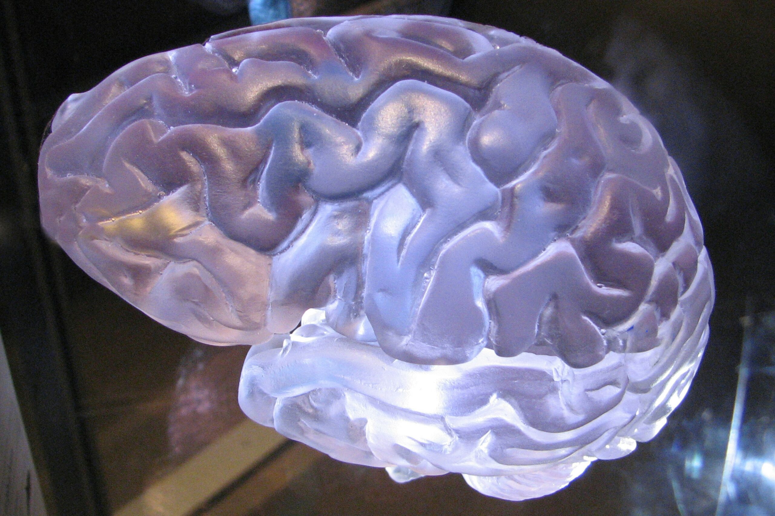
You’ve probably never stopped to truly consider the marvel that happens every second of your waking life: your brain’s incredible ability to interpret the world through light. It’s a silent, seamless process, so automatic that you might take it for granted. But behind your everyday experience of seeing, there’s a sophisticated biological system, a symphony of specialized cells and intricate neural pathways working in concert. This article will take you on a journey, demystifying how your brain transforms fleeting photons into the rich tapestry of images that define your reality.
Before your brain even gets involved, the first crucial step is light’s entry into your visual system. Imagine yourself stepping out into a brightly lit room. The instant photons, those tiny packets of energy, bombard your eyes. This isn’t a passive reception; your eye is an exquisitely designed organ, primed to capture and begin processing this incoming information.
The Cornea: The First Lens
The journey of light begins with the cornea, the transparent, dome-shaped outer layer at the front of your eye. Think of it as your eye’s primary lens. Its curved surface is responsible for bending, or refracting, a significant portion of the incoming light. This bending is essential; it focuses the light rays, ensuring they converge towards the back of your eye, where the magic of vision truly begins. The cornea’s transparency is vital, allowing light to pass through unimpeded, and its smooth surface is critical for sharp image formation. Any irregularities here, like those experienced with astigmatism, can lead to blurred vision because the light isn’t being refracted evenly.
The Pupil and Iris: Controlling the Illumination
Following the cornea, light encounters the pupil, the dark, circular opening in the center of your iris. The iris, that colored part of your eye you might admire in others, acts like the diaphragm of a camera. It’s a muscular structure that controls the size of the pupil. In bright light, your iris constricts the pupil, making it smaller. This reduces the amount of light entering your eye, preventing overstimulation of the sensitive photoreceptor cells at the back. Conversely, in dim light, your iris relaxes, dilating the pupil to allow more light in, maximizing your ability to see in low-light conditions. This automatic adjustment is a testament to your body’s efficient regulation of sensory input.
The Lens: Fine-Tuning the Focus
After passing through the pupil, light strikes the lens, a flexible, transparent structure situated behind the iris. Unlike the cornea, which provides a fixed refractive power, the lens can change its shape. This remarkable ability, called accommodation, allows you to focus on objects at different distances. When you look at something far away, the ciliary muscles surrounding the lens relax, making the lens flatter. When you shift your gaze to a nearby object, these muscles contract, causing the lens to become more curved. This fine-tuning of focus ensures that light from objects both near and far is precisely directed onto the retina.
The Vitreous Humor: Maintaining the Eye’s Shape
The bulk of your eyeball is filled with a gel-like substance called the vitreous humor. While it doesn’t actively refract light, it plays a crucial role in maintaining the eye’s spherical shape. This structural integrity is vital for the optical system to function correctly, ensuring that the light rays are directed onto the retina in the proper manner. Occasionally, you might notice small specks or floaters drifting across your vision. These are often tiny clumps within the vitreous humor that cast shadows on the retina.
Understanding how the brain interprets light is a fascinating topic that delves into the complexities of human perception. For those interested in exploring this subject further, a related article can be found at Freaky Science, which discusses the intricate processes involved in visual perception and how our brains decode the various wavelengths of light to create the images we see. This exploration not only highlights the remarkable capabilities of our sensory systems but also sheds light on the underlying mechanisms that govern our experience of the world around us.
The Retina: Where Light Becomes Neural Signals
The retina, a thin layer of tissue lining the back of your eye, is where the real transformation occurs. This is where light energy is converted into electrical signals that your brain can understand. It’s a complex neural tissue, packed with specialized cells that are highly sensitive to light.
Photoreceptors: The Light Detectors
The stars of the retinal show are the photoreceptor cells: rods and cones. You have millions of these cells, each designed for a specific role in capturing light.
Rods: Masters of Low Light
Scattered throughout the retina, but concentrated in the periphery, are your rods. These are your low-light specialists, responsible for your ability to see in dim conditions and for peripheral vision. While they are incredibly sensitive to light, rods don’t differentiate between colors. This is why in twilight or moonlight, you primarily see in shades of gray. Rods contain a photopigment called rhodopsin, which undergoes a chemical change when it absorbs photons. This change initiates a cascade of events that ultimately leads to an electrical signal.
Cones: The Color and Detail Enthusiasts
Clustered in the central part of the retina, particularly in a region called the fovea, are your cones. These are your daytime, color, and fine detail detectors. You have three types of cones, each sensitive to different wavelengths of light: red, green, and blue. By comparing the signals from these different cone types, your brain can perceive the entire spectrum of colors. The fovea, with its high density of cones, is your area of sharpest vision, essential for reading, recognizing faces, and other detailed visual tasks.
The Interplay of Neurons: From Photoreceptors to the Optic Nerve
The photoreceptors don’t communicate directly with the optic nerve. Instead, they synapse with a layer of intermediate neurons within the retina. These include bipolar cells and amacrine cells. Bipolar cells act as intermediaries, collecting signals from multiple photoreceptors and relaying them to the ganglion cells. Amacrine cells are involved in more complex processing, modulating the signals and contributing to tasks like edge detection.
Bipolar Cells: The Relay Stations
Bipolar cells are crucial for transmitting the signals generated by rods and cones. There are different types of bipolar cells, some of which are excited by light (ON bipolar cells) and others that are inhibited by light (OFF bipolar cells). This differential response contributes to the initial processing of contrast and brightness.
Ganglion Cells: The Output Neurons
The final layer of neurons in the retina before the optic nerve is the ganglion cells. These cells receive input from the bipolar cells and generate action potentials – the electrical impulses that will travel to the brain. Each ganglion cell receives input from a specific receptive field, a small area of the retina. The pattern of firing of these ganglion cells encodes information about the light intensity and color within their receptive fields.
The Journey to the Brain: The Optic Nerve and Beyond

The axons of the ganglion cells bundle together to form the optic nerve, a thick cable that exits the back of your eye. This nerve is your visual highway, carrying all the information from your retina to your brain.
The Optic Chiasm: A Crucial Crossroads
As the optic nerves from both eyes travel towards the brain, they meet at a critical junction called the optic chiasm. Here, a remarkable crossover occurs: information from the nasal (inner) half of each retina crosses over to the opposite side of the brain, while information from the temporal (outer) half remains on the same side. This means that your left hemisphere receives visual information from the right visual field (which encompasses input from the nasal half of the left retina and the temporal half of the right retina), and your right hemisphere receives information from the left visual field. This crossover is essential for integrating visual information from both eyes into a coherent perception of the world.
The Lateral Geniculate Nucleus (LGN): The Thalamic Relay
After the optic chiasm, the visual pathway continues to the lateral geniculate nucleus (LGN), a structure located within the thalamus. The LGN acts as a crucial relay station, receiving input from the optic nerve and further processing and organizing this visual information before sending it on to the visual cortex. The LGN has several layers, with different layers receiving input from different types of ganglion cells (e.g., those carrying information about form and movement versus those carrying color information). This layering allows for a preliminary segregation of visual features.
The Superior Colliculus: Guiding Eye Movements
A smaller portion of the optic nerve fibers also projects to the superior colliculus, a structure in the midbrain. The superior colliculus plays a vital role in controlling eye movements, helping you to quickly orient your gaze towards interesting visual stimuli. It’s like an automatic reflex system, allowing you to react to sudden movements or important visual cues in your environment.
The Visual Cortex: Where Meaning is Made

The majority of visual information, after passing through the LGN, arrives at the primary visual cortex (V1), also known as the striate cortex, located at the back of your brain in the occipital lobe. This is where the raw electrical signals from your eyes are transformed into the conscious perception of vision.
Primary Visual Cortex (V1): The Foundation of Vision
V1 is the initial processing center for visual information. Neurons in V1 are organized in a retinotopic map, meaning that adjacent areas of the retina are represented by adjacent areas in the cortex. This map is not perfectly preserved but provides a spatial representation of the visual field. Neurons in V1 are highly specialized, responding to specific features of the visual input, such as the orientation of lines, edges, and simple patterns.
Orientation Selectivity: Detecting Lines and Edges
One of the most fundamental discoveries in visual neuroscience was that neurons in V1 are selective for the orientation of lines. Some neurons will fire strongly when they detect a vertical line, while others will respond best to horizontal or diagonal lines. This fundamental feature detection is crucial for building up more complex visual representations.
Ocular Dominance Columns: Input from Each Eye
V1 also exhibits ocular dominance columns, which are regions where neurons preferentially respond to input from either the left or the right eye. This organization reflects the input from the LGN and is a step towards binocular vision, where information from both eyes is combined.
Simple and Complex Cells: Building Blocks of Perception
Within V1, researchers have identified two main types of cells: simple cells and complex cells. Simple cells respond best to lines or edges of a specific orientation at a precise location in their receptive field. Complex cells, on the other hand, respond to lines or edges of a specific orientation anywhere within their receptive field, and many are also sensitive to the direction of movement. These cells act as the building blocks for more sophisticated visual processing.
Beyond V1: The Extrastriate Visual Areas
From V1, visual information fans out to a network of interconnected extrastriate visual areas (V2, V3, V4, V5, etc.). Each of these areas specializes in processing different aspects of the visual scene, gradually building a richer and more complex understanding of what you are seeing.
V2: Processing Contours and Depth
V2 receives input from V1 and begins to process more complex visual features, such as illusory contours (lines that appear to exist even though they are not physically drawn) and information related to depth perception.
V4: Color and Form Perception
Area V4 is critically involved in processing color and form. Neurons in V4 are highly tuned to different colors and are crucial for your ability to perceive the consistent color of an object even under varying lighting conditions. It also plays a role in recognizing complex shapes.
V5 (MT): Motion Perception
Area V5, also known as the middle temporal area (MT), is the brain’s dedicated motion processing center. Neurons in V5 are highly sensitive to the speed and direction of movement. Damage to V5 can result in akinetopsia, a condition where individuals are unable to perceive motion, seeing the world as a series of static snapshots.
Understanding how the brain interprets light is a fascinating topic that delves into the complexities of human perception. Recent studies have shown that our brains not only process light but also assign meaning to the colors and brightness we see, influencing our emotions and behaviors. For those interested in exploring this subject further, an insightful article can be found at Freaky Science, which discusses the intricate relationship between light perception and cognitive functions. This connection between light and our mental processes highlights the remarkable capabilities of the human brain.
The Brain as an Interpreter: Constructing Your Visual Reality
| Aspect | Information |
|---|---|
| Photoreceptors | Specialized cells in the retina that convert light into electrical signals |
| Retinal Ganglion Cells | Receive signals from photoreceptors and transmit them to the brain via the optic nerve |
| Visual Cortex | Part of the brain that processes and interprets visual information received from the eyes |
| Color Perception | Brain interprets different wavelengths of light as different colors |
| Light Intensity | Brain adjusts sensitivity to light levels through pupil dilation and neural adaptation |
It’s important to understand that your brain doesn’t passively receive images like a camera. Instead, it actively constructs your visual reality based on the incoming sensory data, your past experiences, expectations, and even your current emotional state.
Top-Down Processing: The Influence of Expectations
While the journey from the eye to the brain is often described as “bottom-up” processing (starting with sensory input), your brain also engages in “top-down” processing. This means that your prior knowledge, memories, and expectations can influence how you interpret visual information. For example, if you expect to see a familiar face in a crowd, your brain might be more likely to “fill in the gaps” and recognize that face, even if the visual information is not perfectly clear. This is why optical illusions can be so compelling; they exploit the way your brain makes assumptions about the visual world.
Object Recognition: Putting the Pieces Together
As visual information flows through the extrastriate cortex, different pathways become engaged. The ventral stream, often called the “what” pathway, projects from V1 down to the temporal lobe and is crucial for object recognition. This pathway allows you to identify what you are seeing – a chair, a tree, a person. It involves processing features like shape, color, and texture to form a complete representation of an object.
Spatial Processing: Navigating Your World
The dorsal stream, often referred to as the “where” or “how” pathway, projects from V1 up to the parietal lobe. This pathway is responsible for processing spatial information, including the location of objects in space, their movement, and how you interact with them. This is essential for navigation, reaching for objects, and understanding the relationships between different elements in your environment.
The Role of Memory and Emotion
Your memories significantly influence your visual perception. A familiar scent might trigger a visual memory of a place, and your emotional state can color how you perceive visual cues. For instance, if you are feeling anxious, you might be more likely to interpret ambiguous visual stimuli as threatening. This demonstrates the interconnectedness of our sensory systems and cognitive processes.
By understanding how your brain processes light, you gain a deeper appreciation for the complexity and elegance of your own visual system. It’s a constant dance between the external world and your internal interpretation, a remarkable feat of biological engineering that allows you to experience the vibrant, detailed, and ever-changing world around you. Every glance, every image, is a testament to this extraordinary process.

Why Your Brain Had to Invent Magenta
FAQs
1. How does the brain interpret light?
The brain interprets light through a complex process involving the eyes, optic nerves, and various regions of the brain, including the visual cortex. Light enters the eyes and is converted into electrical signals that are sent to the brain for processing.
2. What role do photoreceptor cells play in the interpretation of light by the brain?
Photoreceptor cells, specifically rods and cones, are located in the retina of the eye and are responsible for detecting light. Rods are sensitive to low light levels and are important for night vision, while cones are responsible for color vision and detail in bright light.
3. How does the brain process different colors of light?
The brain processes different colors of light through the stimulation of different types of cones in the retina. There are three types of cones, each sensitive to different wavelengths of light corresponding to red, green, and blue. The brain combines the signals from these cones to perceive a wide range of colors.
4. What is the role of the visual cortex in interpreting light?
The visual cortex, located at the back of the brain, is responsible for processing visual information received from the eyes. It plays a crucial role in interpreting light, recognizing shapes, colors, and motion, and forming the basis for visual perception.
5. How does the brain interpret changes in light intensity and brightness?
The brain interprets changes in light intensity and brightness through the regulation of the pupil size and the adaptation of the visual system to different lighting conditions. This allows the brain to adjust to varying levels of light and maintain visual perception.