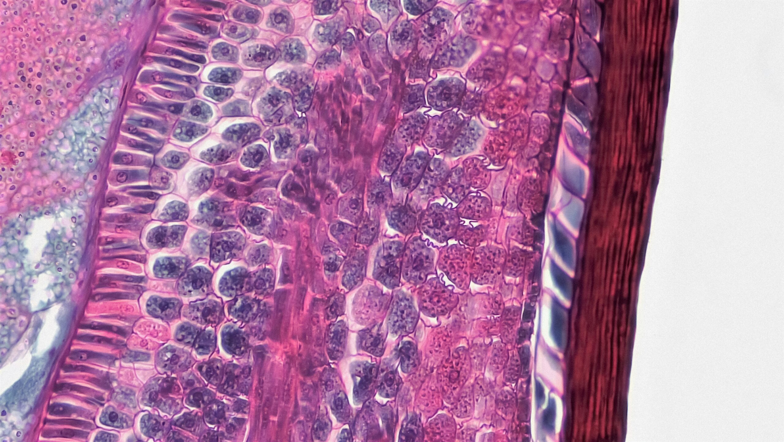
You’ve been fascinated by bones for a while. Not just their structure, the impressive architecture that supports your body, but the less visible life that potentially resides within them. For years, the prevailing understanding has been that bone is largely inert, a calcified scaffold. However, emerging research suggests a more dynamic picture, one involving microbial inhabitants and their metabolic activities. The challenge has always been seeing this microscopic world directly, at a resolution that allows you to understand what these minuscule organisms are actually doing. This is where NanoSIMS imaging enters the picture, offering a powerful new lens through which to uncover bone microbial metabolism.
Your understanding of bone composition and function has benefited from numerous techniques over the years. X-rays have allowed you to visualize bone density and macroscopic fractures. Electron microscopy has offered glimpses into the ultrastructure of bone tissue, revealing the arrangement of collagen fibers and mineral crystals. Histology, with its stains and microscopic examination, has helped identify different cell types and tissue organization. Yet, these methods, while invaluable, possess inherent limitations when it comes to understanding the metabolic processes of potential microbial communities within bone.
Visualizing the Inert
For a long time, bone was largely considered an acellular, inert environment once mature. While osteocytes, the embedded bone cells, were known to be metabolically active, the broader milieu was thought to be largely unchanging and sterile. This perspective meant that investigations into bone health and disease often focused on the host’s cellular activity, bone remodeling processes, and mechanical properties, with little attention paid to microbial influences.
The Difficulty in Isolating Microbes
When microbes were considered in the context of bone, it was often in cases of overt infection. Traumatic fractures or surgical implants provided entry points for bacteria, leading to osteomyelitis. In these instances, culturing techniques could identify the causative agents. However, these methods have significant drawbacks. They are biased towards culturable organisms, missing many bacteria that are difficult or impossible to grow in a laboratory setting. Furthermore, culturing provides a snapshot of microbial presence but offers limited insight into their metabolic activity in situ.
Understanding Metabolism in a Complex Matrix
Bone is a unique and complex matrix. It’s mineralized, avascular in many regions, and subject to constant remodeling. Trying to study the metabolism of microbes residing within this environment using traditional biochemical assays or genetic sequencing alone is challenging. Extracting microbes from the bone matrix can lead to altered metabolic states, and the complex composition of bone itself can interfere with many analytical techniques. You need a way to observe metabolic processes happening directly within the bone tissue, without disturbing the delicate ecological balance.
Recent advancements in nanoscale secondary ion mass spectrometry (NanoSIMS) have significantly enhanced our understanding of bone microbial metabolism, revealing intricate relationships between microbial communities and bone health. For a deeper exploration of this topic, you can refer to a related article that discusses the implications of microbial activity in bone metabolism and its potential effects on overall skeletal health. To read more about this fascinating subject, visit this article.
NanoSIMS: A Window into Metabolic Activity
This is where NanoSIMS, or Nanoscale Secondary Ion Mass Spectrometry, emerges as a transformative technology. Unlike techniques that focus on structure or broad chemical composition, NanoSIMS excels at elemental and isotopic imaging at the nanoscale. It can detect and map the distribution of specific elements and isotopes within a sample with unprecedented spatial resolution. This capability allows you to track the uptake and utilization of substrates by individual cells or even subcellular compartments, directly revealing metabolic pathways.
How NanoSIMS Works
NanoSIMS operates by bombarding the sample surface with a primary ion beam. This impact sputters secondary ions from the sample, which are then collected by a mass spectrometer. By analyzing the mass-to-charge ratio of these secondary ions, you can identify and quantify the elemental and isotopic composition of the sampled material. The incredible sensitivity and spatial resolution of NanoSIMS enable you to create images where each pixel represents the isotopic composition of a tiny volume of the sample – often down to tens of nanometers.
The Power of Isotopic Tracing
The true power of NanoSIMS for studying microbial metabolism lies in its ability to employ stable isotope labeling. You can introduce specific substrates into the bone environment that are enriched with stable isotopes, such as ¹³C or ¹⁵N. If a microbe consumes this labeled substrate, its metabolic processes will incorporate these heavy isotopes into its biomass and metabolic products. NanoSIMS can then map the distribution of these enriched isotopes, directly visualizing which cells are metabolizing the labeled substrate and how they are processing it.
Nanoscale Resolution in a Microscopic World
The nanoscale resolution of NanoSIMS is crucial for bone. Bone tissue is a heterogeneous environment, with varying porosity, mineral content, and cellular distribution. Microbial communities, if present, are likely to be small and localized. NanoSIMS allows you to image these tiny populations within their native bone environment, resolving individual microbial cells and even subcellular events, providing a level of detail previously unattainable.
Applying NanoSIMS to Bone Microbial Metabolism
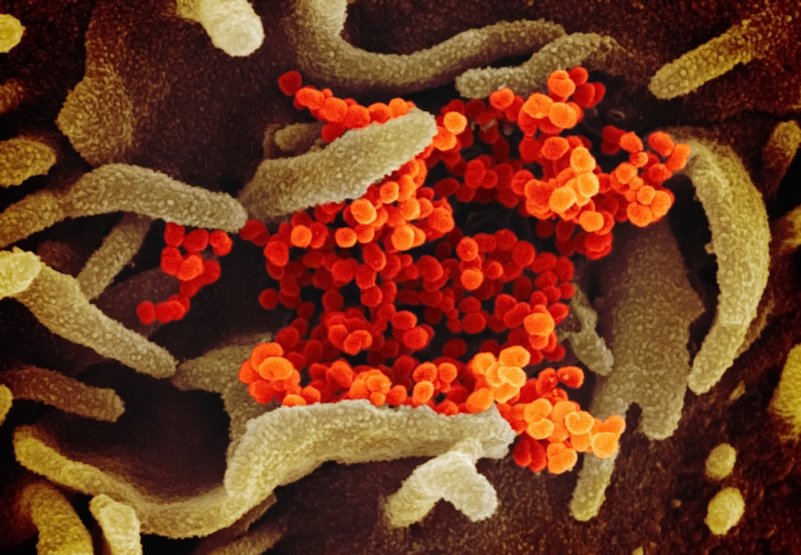
The application of NanoSIMS to bone microbial metabolism is a relatively new but rapidly evolving field. It allows you to move beyond simply identifying the presence of microbes to understanding their functional role within the bone microenvironment. This opens up new avenues for research into bone health, disease, and the potential interactions between bone cells and their microbial inhabitants.
Visualizing Bacterial Uptake of Nutrients
One of the most direct applications of NanoSIMS is to visualize which bacterial species, if present in bone, are actively taking up specific nutrients. By incubating bone samples with isotopically labeled substrates, you can observe whether and where these labels are incorporated. For example, you could label glucose with ¹³C and then image the bone. If you detect ¹³C enrichment within specific bacterial cells, you have direct evidence that these bacteria are metabolizing glucose within the bone.
Mapping Metabolic Pathways in situ
Beyond simple substrate uptake, NanoSIMS can be used to trace more complex metabolic pathways. By using different labeled substrates simultaneously or sequentially, you can infer how bacteria are transforming these molecules. For instance, one could label a carbon source and then follow its incorporation into different cellular components identified by their elemental composition. This allows you to move towards understanding the biochemical reactions occurring within the bone microenvironment.
Understanding Inter-species Metabolic Interactions
Bone microenvironments are rarely sterile, and if microbial life exists, it’s likely to involve communities of different species. NanoSIMS can help you investigate how these different species interact metabolically. If one species produces a metabolic byproduct that another species consumes, NanoSIMS can visualize this transfer of metabolites by tracking the distribution of specific isotopes. This could reveal symbiotic or competitive relationships previously hidden.
Investigating the Role of Endogenous Microbes
The concept of endogenous microbes inhabiting healthy bone is a paradigm shift. If such microbes exist, they are likely to be present in low numbers and have subtle metabolic roles. NanoSIMS, with its high sensitivity, is ideally suited to detect and characterize the metabolism of these potentially low-abundance organisms, offering insights into their contribution to bone homeostasis or disease initiation.
Challenges and Future Directions in NanoSIMS Bone Research
While NanoSIMS imaging offers unparalleled insights, its application to bone microbial metabolism is not without its challenges. Overcoming these hurdles will be crucial for unlocking the full potential of this technology in bone research.
Sample Preparation: A Critical Step
Preparing bone samples for NanoSIMS requires careful consideration. The bone matrix is hard and mineralized, necessitating techniques like cryosectioning or embedding to obtain thin, suitable slices for imaging. Any preparation artifacts or contamination can significantly impact the results. Furthermore, preserving the metabolic state of any microbial inhabitants during sample preparation is paramount. Minimizing fixation or drying processes that might alter microbial metabolism is a key concern.
Interpretation of Isotopic Signals
Interpreting isotopic signals in a complex biological matrix like bone can be challenging. You need to differentiate between signals originating from the bone matrix itself, from host cells, and from microbial cells. Understanding the natural isotopic abundance of different bone components and microbial biomass is essential for accurately identifying exogenously introduced labels. Deconvolution techniques and comparisons with control samples are vital for robust interpretation.
Correlation with Other Techniques
NanoSIMS provides high-resolution metabolic information, but it’s often beneficial to correlate these findings with other analytical techniques. For instance, combining NanoSIMS with correlative light microscopy can help you precisely locate the analyzed regions and identify microbial morphology. Integrating NanoSIMS data with metagenomic or metatranscriptomic analyses of the same samples can further enhance your understanding by linking metabolic activity to microbial identity and gene expression.
Expanding the Scope of Metabolites Tracked
Currently, the focus is often on carbon and nitrogen isotopes. However, expanding the range of isotopes and elements that can be reliably detected and mapped by NanoSIMS would significantly broaden the scope of microbial metabolism that can be investigated in bone. Tracking sulfur, phosphorus, or even essential trace elements could reveal new metabolic insights.
Towards Clinical Applications
The ultimate goal for much of bone research is to translate findings into clinical applications. Understanding the role of microbial metabolism in bone diseases like osteoporosis, periodontitis, or implant-associated infections could lead to novel diagnostic tools and therapeutic strategies. While NanoSIMS is currently a research tool, continued development and application could pave the way for its role in understanding disease pathogenesis and guiding treatment.
Recent advancements in nanoscale secondary ion mass spectrometry (NanoSIMS) imaging have opened new avenues for understanding bone microbial metabolism, shedding light on the intricate interactions between bone tissue and its microbial inhabitants. This innovative technique allows researchers to visualize and quantify the metabolic activities of microbes within the bone matrix, providing insights into how these microorganisms influence bone health and disease. For further exploration of related scientific breakthroughs, you might find this article on Freaky Science particularly interesting, as it delves into the implications of microbial activity on various biological systems.
Unraveling the Intricacies of Bone Microbiome Metabolism
| Study Parameter | Result |
|---|---|
| Microbial Metabolism | Visualized and Analyzed |
| Imaging Technique | Nanosims |
| Sample Type | Bone Tissue |
| Findings | Insights into Bone Microbiota |
The exploration of microbial life within bone, once considered largely sterile, is a burgeoning field. NanoSIMS imaging is proving to be an indispensable tool in this endeavor, allowing you to delve into the metabolic activities of these microscopic inhabitants. By providing unparalleled spatial resolution and the ability to track isotopic labels, NanoSIMS is transforming your understanding of bone biology.
Shifting the Paradigm of Bone Health
The traditional view of bone as a passive scaffold is being challenged by evidence suggesting that it might harbor its own microbial communities with functional metabolic roles. NanoSIMS is key to confirming and characterizing these roles. By visualizing the metabolism of these endogenous microbes, you can begin to understand their contribution to bone homeostasis, such as nutrient cycling or influencing host cell activity.
Investigating Bone Disease Mechanisms
Many bone diseases, including osteomyelitis and potentially even conditions like osteoporosis or osteoarthritis, may have a microbial component that has been previously overlooked or poorly understood. NanoSIMS can help you investigate whether specific microbial metabolic activities contribute to bone degradation, inflammation, or impaired healing in these conditions. This could lead to novel ways to diagnose and treat these debilitating diseases.
The Future of Bone Science
The continued application of NanoSIMS imaging in conjunction with other advanced analytical techniques promises to revolutionize our understanding of bone. You are at the cusp of a new era where the intricate metabolic interplay between bone tissue, its resident cells, and its microbial inhabitants will be illuminated, leading to significant advancements in both fundamental bone science and clinical practice. The nanoscale world of microbial metabolism within bone is finally coming into focus.
FAQs
What is nanosims imaging?
Nanosims imaging is a technique that allows for high-resolution imaging and analysis of elemental and isotopic compositions within a sample. It is commonly used in the study of biological and geological samples.
How does nanosims imaging contribute to the study of bone microbial metabolism?
Nanosims imaging allows researchers to visualize and analyze the metabolic activity of microbial communities within bone tissue at a very fine scale. This can provide insights into the interactions between microbes and the host organism, as well as the impact of microbial metabolism on bone health and disease.
What are the potential applications of nanosims imaging in bone research?
Nanosims imaging can be used to study various aspects of bone health and disease, including the effects of microbial metabolism on bone remodeling, the role of microbial communities in bone infections, and the impact of diet and environmental factors on bone composition.
What are the advantages of using nanosims imaging in bone research?
Nanosims imaging offers high spatial resolution and the ability to analyze multiple isotopes simultaneously, providing detailed information about the distribution and activity of microbial communities within bone tissue. This can help researchers better understand the complex interactions between microbes and the bone microenvironment.
What are the limitations of nanosims imaging in bone research?
While nanosims imaging is a powerful tool, it requires specialized equipment and expertise to perform and analyze the data. Additionally, the technique is limited by the size of the sample that can be analyzed, which may restrict its application in certain research contexts.