The p53 protein, a vital component of cellular machinery, plays a critical role in maintaining genomic integrity. Its multifaceted functions, primarily centered on preventing the propagation of damaged DNA, have led to its designation as “guardian of the genome.” This article will explore the intricate mechanisms by which p53 operates, its regulation, and the profound implications of its dysfunction in human health.
The p53 gene, known officially as TP53, is one of the most extensively studied genes in molecular biology. It acts as a tumor suppressor, meaning its normal function is to inhibit the development of cancer. The TP53 gene encodes a protein, also referred to as p53, which is a transcription factor. As a transcription factor, p53 binds to specific DNA sequences and influences the expression of other genes, thereby orchestrating a complex cellular response to stress.
Discovery and Early Characterization of p53
The p53 protein was initially identified in the late 1970s and early 1980s. Early research characterized it as a protein that was frequently overexpressed in various tumors. This observation led some initial researchers to wrongly classify p53 as an oncogene, a gene that promotes cancer. However, further investigation revealed a more nuanced and critical role. It was discovered that mutations in the TP53 gene were exceptionally common in human cancers, occurring in over 50% of all malignancies. This high mutation frequency underscored its importance as a suppressor of tumorigenesis. The realization that loss-of-function or dominant-negative mutations in TP53 allowed cells with damaged DNA to proliferate and accumulate further genetic alterations was a pivotal moment in understanding cancer biology.
The p53 Family: Related Proteins with Similar Functions
The p53 protein is not an isolated entity but rather belongs to a family of related proteins that share structural and functional similarities. This family, often referred to as the p53 family or the DNA-binding protein (DBP) family, includes p63 and p73. While p53 is ubiquitously expressed and plays the primary role in genomic surveillance in most cells, p63 and p73 exhibit more restricted expression patterns, often found in specific tissues and during development. Despite these differences, they can also function as transcription factors, regulating gene expression and participating in cellular processes like cell differentiation, apoptosis, and stress response. However, their roles are generally considered to be distinct from, though sometimes overlapping with, the canonical functions of p53.
The p53 protein, often referred to as the “guardian of the genome,” plays a crucial role in maintaining cellular integrity by regulating the cell cycle and preventing genomic mutations. For a deeper understanding of its functions and implications in cancer biology, you can explore a related article that delves into the mechanisms by which p53 exerts its protective effects on the genome. To read more about this fascinating topic, visit this article.
The Mechanistic Actions of p53
The “guardian” role of p53 stems from its ability to act as a central sensor of cellular stress. Upon detecting various forms of cellular damage or oncogenic signals, p53 is activated and initiates a cascade of downstream events aimed at either repairing the damage or eliminating the compromised cell. This intricate regulatory network involves protein-protein interactions, post-translational modifications, and the transcriptional activation of a diverse array of target genes.
Sensing Cellular Stress: Triggers for p53 Activation
A variety of cellular insults can trigger the activation of p53. These stressors act through distinct signaling pathways, ultimately converging on p53 stabilization and transcriptional activity.
DNA Damage.
One of the most potent activators of p53 is the presence of DNA damage. This can arise from various sources, including ionizing radiation, UV radiation, chemical mutagens, and errors during DNA replication. The cellular machinery responsible for detecting DNA breaks, such as the ataxia-telangiectasia mutated (ATM) kinase and the ataxia-telangiectasia and rad3-related (ATR) kinase, plays a crucial role in this process. Upon sensing DNA damage, ATM and ATR phosphorylate p53 at specific serine residues, most notably serine 15. This phosphorylation event is critical for stabilizing p53 and preventing its degradation by cellular proteases.
Oncogene Activation.
The inappropriate activation of oncogenes, a hallmark of cancer development, can also induce p53 activation. This phenomenon is known as oncogene-induced senescence. Oncogenes such as MYC, RAS, and E2F can promote aberrant cell proliferation and disrupt normal cellular homeostasis. In response to this aggressive growth signaling, p53 is activated, leading to cell cycle arrest or apoptosis, thereby preventing the formation of a tumor. This indicates that p53 not only responds to damage but also to precancerous signals.
Hypoxia and Nutrient Deprivation.
Low oxygen levels (hypoxia) and lack of essential nutrients can also trigger p53-mediated responses. Hypoxia, for example, can activate signaling pathways that ultimately lead to p53 stabilization. Similarly, metabolic stress, such as glucose deprivation, can induce p53 activation. These responses aim to conserve cellular resources, limit cell growth, and promote survival in challenging environments, or to initiate self-destruction if the stress is too severe to overcome.
p53 as a Transcription Factor: Orchestrating Cellular Responses
Once activated and stabilized, p53 translocates to the nucleus, where it binds to specific DNA sequences called p53 response elements (p53REs). These p53REs are typically located in the promoter regions of target genes, and their recognition by p53 leads to either the activation or repression of gene transcription. The specific outcome depends on the context and the binding partners of p53.
Induction of Cell Cycle Arrest.
A primary function of activated p53 is to halt the cell cycle, providing an opportunity for DNA repair. p53 achieves this by upregulating the expression of genes such as CDKN1A (encoding p21), which inhibits cyclin-dependent kinases (CDKs). CDKs are essential for driving the cell cycle progression through its various phases. By inhibiting CDKs, p53 effectively acts as a brake, preventing the cell from replicating its damaged DNA and passing it on to daughter cells. This arrest can occur at the G1/S transition (preventing entry into DNA synthesis) or at the G2/M transition (preventing entry into mitosis).
Promotion of Apoptosis.
If the DNA damage is too extensive or irreparable, p53 can initiate apoptosis, or programmed cell death. This is a crucial mechanism for eliminating cells that pose a risk to the organism. p53 achieves this by inducing the expression of pro-apoptotic genes, such as BAX and PUMA. These proteins promote the release of cytochrome c from the mitochondria, which in turn activates caspases, the executioner enzymes of apoptosis. The controlled dismantling and removal of the damaged cell prevent the accumulation of mutations that could lead to cancer.
Activation of DNA Repair Pathways.
In situations where DNA damage is potentially repairable, p53 can also promote the expression of genes involved in DNA repair mechanisms. This can include genes that encode enzymes responsible for base excision repair, nucleotide excision repair, and homologous recombination. By upregulating these pathways, p53 facilitates the accurate restoration of the DNA sequence, thus mitigating the risk of permanent mutations.
Regulation of p53 Activity: A Tight Control System
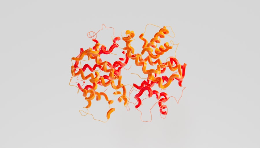
The critical role of p53 necessitates strict control over its activity. Its levels and function are tightly regulated to ensure that it is only activated in response to genuine cellular stress and does not inappropriately interfere with normal cellular processes. This regulation occurs through various mechanisms, primarily involving its stability and transcriptional activity.
Proteasomal Degradation: The MDM2 Pathway
In unstressed cells, p53 levels are kept very low due to a negative feedback loop involving the murine double minute 2 (MDM2) protein. MDM2 is a ubiquitin ligase that binds to p53 and targets it for degradation by the 26S proteasome. This constant turnover ensures that p53 does not accumulate and remain active under normal conditions.
The Role of MDM2 in p53 Stability.
MDM2 binds to the N-terminus of p53 and catalyzes the attachment of ubiquitin chains, marking p53 for destruction. This interaction is crucial. Under normal circumstances, without any cellular stress, p53 is continuously synthesized and degraded by this MDM2-mediated pathway.
p53-Mediated MDM2 Transcription.
Interestingly, p53 itself can also regulate the transcription of the MDM2 gene. When p53 is activated and binds to the MDM2 promoter, it increases the expression of MDM2. This creates a negative feedback loop: as p53 levels rise, it induces more MDM2, which then promotes p53 degradation, thus keeping p53 levels in check. This intricate loop is essential for maintaining cellular homeostasis.
Post-Translational Modifications: Fine-Tuning p53 Function
Beyond its regulation by MDM2, p53 activity is also modulated by a wide array of post-translational modifications (PTMs). These modifications can alter p53’s stability, localization, DNA-binding affinity, and interaction with other proteins.
Phosphorylation.
As mentioned earlier, phosphorylation of p53 at specific serine and threonine residues, such as Ser15, is a key event in its activation. This can be mediated by kinases like ATM, ATR, and DNA-PK. Phosphorylation can disrupt the interaction between p53 and MDM2, leading to p53 stabilization. Other phosphorylation sites can influence p53’s transcriptional activity or its ability to interact with co-activators.
Acetylation.
Acetylation, typically occurring at lysine residues, is another important PTM for p53. Enzymes called histone acetyltransferases (HATs) can acetylate p53, often enhancing its transcriptional activity by promoting its binding to DNA and its interaction with transcription co-activators. Conversely, histone deacetylases (HDACs) can remove acetyl groups, leading to decreased p53 activity.
Ubiquitination and Sumoylation.
Ubiquitination, beyond triggering degradation by MDM2, can also play other roles. For example, some forms of ubiquitination can stabilize p53 or modulate its interactions. Sumoylation, another type of protein modification, can also influence p53’s transcriptional activity and localization. The interplay between these diverse modifications creates a complex regulatory network that fine-tunes p53 response in a context-dependent manner.
The Consequences of p53 Dysfunction: A Gateway to Cancer

Given its central role in preventing genomic instability and uncontrolled cell proliferation, it is no surprise that the inactivation or mutation of p53 is a frequent event in the development of cancer. Loss of functional p53 removes a critical safeguard, allowing cells with accumulated genetic errors to survive and divide, leading to tumor formation and progression.
TP53 Mutations in Cancer: Prevalence and Types
Mutations in the TP53 gene are among the most common genetic alterations found in human cancers, occurring in over half of all sporadic tumors. These mutations can arise through various mechanisms, including point mutations, insertions, deletions, and chromosomal rearrangements.
Missense Mutations.
The most frequent type of TP53 mutation in cancer are missense mutations, which result in the substitution of a single amino acid in the p53 protein. These mutations often occur in the DNA-binding domain of p53, directly impairing its ability to bind to DNA and regulate gene expression. Many of these missense mutations are classified as “gain-of-function” mutations, meaning the mutant p53 protein not only loses its tumor suppressor function but can also acquire new, oncogenic properties, promoting tumor growth and metastasis.
Truncating Mutations.
Truncating mutations, such as nonsense mutations or frameshift mutations, lead to the premature termination of protein synthesis, resulting in a shortened and typically non-functional p53 protein. These mutations effectively eliminate the tumor suppressor activity of p53.
Li-Fraumeni Syndrome: A Hereditary Predisposition to Cancer
Germline mutations in the TP53 gene, meaning mutations present in the egg or sperm cells and thus inherited, lead to a rare but aggressive cancer predisposition syndrome known as Li-Fraumeni Syndrome (LFS). Individuals with LFS have a significantly increased lifetime risk of developing a wide range of cancers, often at early ages.
Inheritance Pattern and Cancer Spectrum.
LFS is inherited in an autosomal dominant pattern, meaning a single copy of the mutated TP53 gene is sufficient to confer a high risk of cancer. Individuals with LFS often develop multiple primary cancers throughout their lifetime, including sarcomas, breast cancer, brain tumors, leukemias, and adrenal cortical carcinomas. The high penetrance and broad spectrum of cancers associated with LFS highlight the indispensable role of p53 in preventing tumorigenesis.
Therapeutic Strategies Targeting p53
The prevalence of TP53 mutations in cancer and the critical nature of p53’s function have made it an attractive target for therapeutic interventions. However, directly restoring the function of mutated p53 has proven challenging. Current research focuses on reactivating mutant p53, inducing apoptosis in p53-deficient cells, or targeting other pathways that might compensate for p53 loss.
Reactivating Mutant p53.
One promising area of research involves developing small molecules that can bind to mutant p53 and restore its wild-type conformation and function. These “p53 reactivators” aim to refold misfolded mutant p53 proteins, enabling them to bind DNA and induce cell cycle arrest or apoptosis.
Exploiting p53-Deficient Tumors.
Another strategy is to exploit the dependency of p53-deficient tumors on other pathways for survival. For instance, some tumors with p53 loss become reliant on other cell cycle regulators. Inhibiting these compensatory pathways can thus selectively kill cancer cells while sparing normal cells. Gene therapy approaches aiming to reintroduce functional p53 into cancer cells are also being explored, although challenges remain in achieving sufficient and sustained expression.
The p53 protein, often referred to as the guardian of the genome, plays a crucial role in maintaining cellular integrity by regulating the cell cycle and preventing the proliferation of damaged DNA. For a deeper understanding of its functions and implications in cancer biology, you might find this article on the topic particularly enlightening. It explores the various mechanisms through which p53 operates and its significance in tumor suppression. To read more about this fascinating protein, you can visit this article.
p53 Beyond the Genome: Emerging Roles and Future Directions
| Property | Value |
|---|---|
| Gene Name | TP53 |
| Function | Tumor Suppressor |
| Cellular Location | Nucleus |
| Role | Regulates cell cycle, apoptosis, and DNA repair |
| Associated Diseases | Cancer, Li-Fraumeni syndrome |
While the role of p53 as a guardian of the genome is well-established, ongoing research continues to uncover new and intriguing functions of this multifaceted protein. These emerging roles highlight the pervasive influence of p53 on cellular physiology and its implications for various biological processes, extending beyond its canonical tumor suppressor activities.
Metabolism and p53 Activity
Recent studies have revealed a significant link between p53 and cellular metabolism. p53 can directly influence metabolic pathways by regulating the expression of genes involved in processes such as glycolysis, oxidative phosphorylation, and fatty acid metabolism. This metabolic control is important for cellular energy production and biosynthesis, and aberrant metabolism is a hallmark of cancer.
Influence on Glycolysis.
p53 can inhibit glycolysis, a major pathway for energy production, especially under stress conditions. By downregulating the expression of key glycolytic enzymes, p53 can help to conserve cellular resources and prevent uncontrolled cell proliferation. This contrasts with the Warburg effect observed in many cancer cells, where glycolysis is upregulated even in the presence of oxygen.
Role in Mitochondrial Function.
p53 also plays a role in regulating mitochondrial function, the primary site of oxidative phosphorylation. It can influence mitochondrial respiration and the production of reactive oxygen species (ROS). Its pro-apoptotic functions are often mediated through interactions with mitochondria, underscoring its control over cellular energy homeostasis and cell death.
Autophagy and p53
Autophagy is a cellular process by which cells degrade and recycle their own components, serving roles in cellular maintenance, stress response, and survival. The relationship between p53 and autophagy is complex and context-dependent.
Dual Nature of p53 in Autophagy.
In some contexts, p53 can induce autophagy, promoting cellular survival under stress. In other situations, particularly when DNA damage is severe, p53 can suppress autophagy, facilitating the execution of apoptosis. This dualistic role suggests that p53 can finely tune the balance between survival and death based on the cellular environment.
Therapeutic Implications of the p53-Autophagy Axis.
Understanding the interplay between p53 and autophagy may offer new therapeutic avenues. For instance, modulating autophagy in p53-deficient tumors could potentially enhance their sensitivity to chemotherapy or other treatments.
The Future of p53 Research
The continued exploration of p53’s diverse functions promises to yield new insights into fundamental biological processes and disease mechanisms. Future research will likely focus on developing more sophisticated tools to investigate p53’s interactions and regulatory networks, with the ultimate goal of translating these discoveries into effective therapeutic strategies for cancer and other diseases. The development of targeted therapies that can either restore p53 function or exploit its absence in cancer cells remains a prominent area of investigation, holding significant potential for improving patient outcomes. Studying the interplay between p53 mutations and the tumor microenvironment will also be crucial for a comprehensive understanding of cancer progression and response to treatment.
FAQs
What is the p53 protein?
The p53 protein is a tumor suppressor protein that plays a crucial role in preventing the development of cancer. It is often referred to as the “guardian of the genome” due to its role in regulating cell division and preventing the proliferation of damaged or abnormal cells.
How does the p53 protein function?
The p53 protein functions by monitoring the integrity of the DNA within cells. When it detects DNA damage or other abnormalities, it can initiate processes that either repair the damage or induce cell death (apoptosis) to prevent the propagation of damaged cells.
What happens when the p53 protein is mutated or dysfunctional?
Mutations or dysfunction in the p53 protein can lead to the unchecked growth and proliferation of damaged or abnormal cells, increasing the risk of cancer development. In fact, mutations in the p53 gene are among the most common genetic alterations in human cancers.
How is the p53 protein regulated?
The activity of the p53 protein is tightly regulated by various cellular mechanisms, including post-translational modifications, protein-protein interactions, and feedback loops. These regulatory mechanisms ensure that the p53 protein is activated only when necessary to maintain genomic stability.
What are the implications of p53 protein research for cancer treatment?
Understanding the role of the p53 protein in cancer development has led to the development of targeted therapies that aim to restore or enhance p53 function in cancer cells. Additionally, p53 status is often used as a prognostic marker in cancer diagnosis and treatment planning.