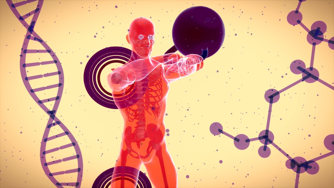
You likely don’t give it much thought, but deep within your body, at this very moment, an extraordinary process is unfolding. It’s a silent, ceaseless marvel, a testament to life’s enduring drive. You are witnessing, though you cannot see it directly, the incredible process of cell creation in the human body. This isn’t just about growth; it’s about repair, renewal, and the very essence of your continued existence. From the moment you were a single cell, this intricate ballet of biological machinery has been orchestrating your form, your function, and your very being.
At the heart of every cell’s ability to create itself, and indeed the ability for all cells to form and function within you, lies an astonishing molecule: Deoxyribonucleic Acid, or DNA. Imagine it as the ultimate instruction manual, a meticulously written code that dictates everything about you, from the color of your eyes to the way your cells communicate. When a cell is poised to reproduce, this blueprint undergoes a remarkable duplication process.
The Double Helix: A Masterpiece of Structure
To understand cell creation, you must first appreciate the structure of DNA. It’s famously depicted as a double helix, a twisted ladder. The sides of this ladder are made of sugar and phosphate molecules, while the rungs are formed by pairs of four chemical bases: Adenine (A), Thymine (T), Guanine (G), and Cytosine (C). The crucial rule of pairing is that A always pairs with T, and G always pairs with C. This specific pairing is fundamental to the entire replication process, ensuring accuracy.
DNA Replication: Copying the Code with Precision
Before any cell can divide, its DNA must be duplicated. This is a highly orchestrated event involving a symphony of enzymes. The process begins with an enzyme called helicase, which acts like a zipper, unwinding the DNA double helix, separating the two strands. Once separated, each strand serves as a template for building a new complementary strand. Enzymes called DNA polymerases then move along each template strand, reading the sequence of bases and adding the corresponding complementary bases (A with T, G with C) to build the new DNA molecule. This is achieved with astonishing speed and incredible accuracy. While errors can occur, the cell has sophisticated proofreading mechanisms to correct them, minimizing the chances of mutations that could lead to problems. The result of this intricate dance is two identical DNA double helices, ready to be passed on to daughter cells.
Genes: The Functional Units of the Code
Within this vast DNA library, specific segments are organized into genes. Each gene holds the instructions for building a particular protein, and proteins are the workhorses of your cells, carrying out a vast array of functions. When a cell needs to create new cells, it essentially needs to duplicate these genetic instructions and the cellular machinery that reads and utilizes them. The DNA replication process ensures that each new cell receives a complete and accurate copy of all the genes, preserving the integrity of your genetic information across generations of cells.
Cells are the fundamental building blocks of the human body, orchestrating a complex symphony of biological processes that contribute to growth, repair, and overall function. Understanding how these cells work together is crucial for grasping the intricacies of human biology. For a deeper dive into this fascinating topic, you can explore the related article on cell functions and their role in forming the human body at this link.
Mitosis: The Symphony of Cell Division for Growth and Repair
Once the DNA has been faithfully duplicated, the cell is ready to divide. For most cell creation in your body – the ones responsible for growth during childhood, replacing worn-out tissues, and healing injuries – the process is mitosis. It’s a meticulously timed and regulated series of events that ensures each new daughter cell receives a complete set of chromosomes.
The Cell Cycle: A Highly Regulated Journey
Mitosis isn’t a spontaneous event; it’s part of a larger, highly regulated cycle called the cell cycle. This cycle has distinct phases:
Interphase: The Preparatory Stage
This is the longest phase of the cell cycle, where the cell grows, carries out its normal functions, and, most importantly, duplicates its DNA. Interphase itself is divided into three sub-phases: G1 (Gap 1), where the cell grows and synthesizes proteins; S (Synthesis), where DNA replication occurs; and G2 (Gap 2), where the cell prepares for division by synthesizing proteins and checking the replicated DNA for errors.
M Phase: The Division Itself
This is the culmination of the cell cycle and includes mitosis (nuclear division) and cytokinesis (cytoplasmic division). Mitosis is further divided into several stages:
Prophase: Condensing the Chromosomes
In this initial stage, the duplicated chromosomes, which have been replicating as loose chromatin, begin to condense and thicken, becoming visible under a microscope. Each chromosome now consists of two identical sister chromatids, joined at a region called the centromere. The nuclear envelope, which encloses the DNA, also starts to break down.
Metaphase: Aligning for Separation
The chromosomes move to the center of the cell, aligning themselves along an imaginary plane called the metaphase plate. Microtubules, which form the mitotic spindle, attach to the centromeres of each chromosome, ensuring proper alignment and preparing for their separation. This precise alignment is critical to prevent errors in chromosome distribution.
Anaphase: Pulling Apart the Duplicates
The sister chromatids are now pulled apart by the shortening of the mitotic spindle microtubules. Each chromatid, now considered an individual chromosome, moves towards opposite poles of the cell. This ensures that each future daughter cell will receive an identical set of chromosomes.
Telophase: Reforming the Nuclei
As the chromosomes reach the opposite poles, they begin to decondense, and new nuclear envelopes form around each set of chromosomes, creating two distinct nuclei.
Cytokinesis: Dividing the Cytoplasm
Following mitosis, the cell’s cytoplasm divides. In animal cells, this involves the formation of a cleavage furrow, a pinching inward of the cell membrane, which eventually divides the cell into two distinct daughter cells, each with its own nucleus and cytoplasm. Plant cells, with their rigid cell walls, form a cell plate that grows outward to divide the cell.
Meiosis: Crafting the Gametes for Reproduction
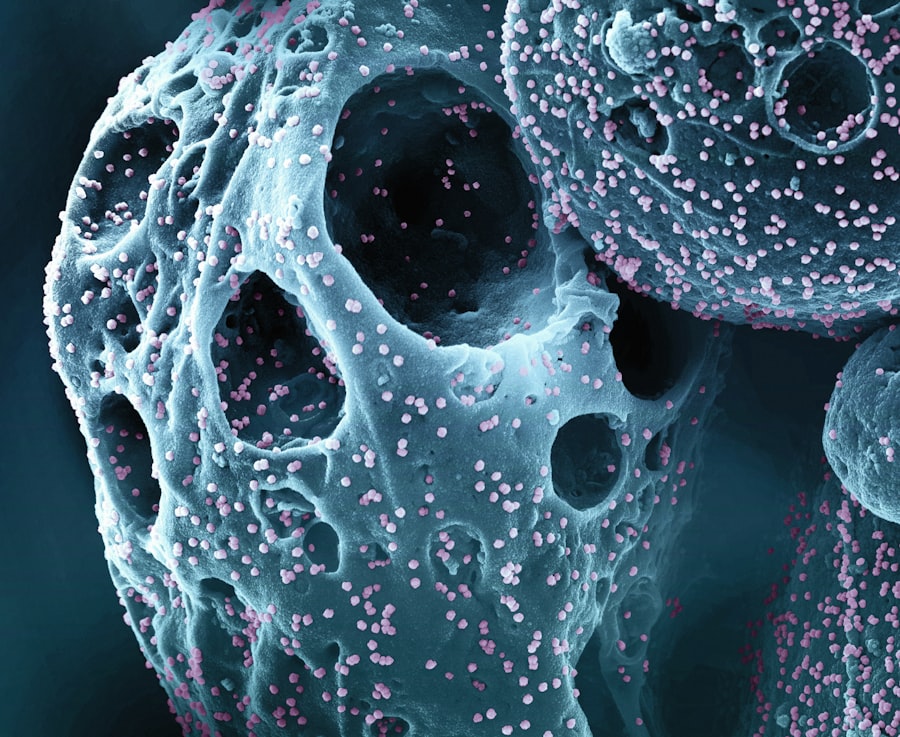
While mitosis is about creating identical copies for growth and repair, there’s another crucial type of cell division: meiosis. This process is specifically designed for sexual reproduction, creating specialized cells called gametes – sperm in males and egg cells in females. The key difference here is that meiosis doesn’t produce identical copies; it produces genetically unique cells with half the number of chromosomes.
The Two Rounds of Division
Meiosis is a more complex process than mitosis, involving two rounds of division rather than one. This two-step process is essential for reducing the chromosome number by half.
Meiosis I: Reducing the Chromosome Number
This is where the magic of halving the chromosome number begins.
Prophase I: The Dance of Homologous Chromosomes
This is a particularly unique and important stage. Homologous chromosomes – pairs of chromosomes, one inherited from each parent – come together and pair up. During this pairing, a process called crossing over can occur. This is where segments of DNA are exchanged between homologous chromosomes, shuffling genetic material and creating new combinations of genes. This is a primary source of genetic variation in sexually reproducing organisms.
Metaphase I: Pairing at the Center
The paired homologous chromosomes, not individual chromosomes as in mitosis, align at the metaphase plate. Their orientation is random, meaning each pair can align in one of two ways, further contributing to genetic diversity.
Anaphase I: Separating the Pairs
The homologous chromosomes are pulled apart to opposite poles of the cell. Importantly, the sister chromatids remain attached. This is the step where the chromosome number is effectively halved.
Telophase I and Cytokinesis: Two Haploid Cells Form
The cell divides into two daughter cells. Each daughter cell now contains one chromosome from each homologous pair, and each chromosome still consists of two sister chromatids. These cells are considered haploid, meaning they have half the number of chromosomes as the original diploid cell.
Meiosis II: Separating the Sister Chromatids
This second round of division is very similar to mitosis.
Prophase II: Preparing for Separation
The chromosomes condense again, and the nuclear envelope breaks down, similar to prophase in mitosis.
Metaphase II: Alignment at the Center
The chromosomes, each still composed of two sister chromatids, align at the metaphase plate in each of the two haploid cells.
Anaphase II: Splitting the Sister Chromatids
The sister chromatids are now pulled apart by the spindle fibers and move to opposite poles of each cell. This effectively separates the duplicated genetic material.
Telophase II and Cytokinesis: Four Haploid Gametes
Each of the two cells from Meiosis I divides again, resulting in a total of four daughter cells. These four cells are haploid and genetically unique, each containing a single set of chromosomes. These are your gametes, ready to combine with a gamete from the opposite sex to form a new diploid individual.
Genetic Diversity: The Power of Meiosis
The processes of crossing over and the independent assortment of homologous chromosomes during meiosis are the cornerstones of genetic diversity. They ensure that each offspring is genetically unique, a crucial factor for the long-term survival and adaptation of species. Without meiosis, all offspring would be genetically identical to their parents, making them highly vulnerable to environmental changes and diseases.
Stem Cells: The Master Architects of Development and Regeneration
You’ve learned about the regular cell creation that fuels your daily life and the specialized creation of gametes. But there’s a third, profoundly important category of cells involved in creation: stem cells. These are your body’s master cells, possessing the unique ability to develop into many different cell types and to self-renew through cell division.
Totipotent Stem Cells: The Ultimate Creators
At the earliest stages of embryonic development, you possess totipotent stem cells. These are the most versatile, capable of differentiating into all cell types of the body, including the placenta. The zygote, the first cell formed after fertilization, is totipotent.
Pluripotent Stem Cells: The Versatile Designers
As development progresses, totipotent cells differentiate into pluripotent stem cells. These cells can differentiate into any cell type in the body but cannot form the placenta. Embryonic stem cells are a prime example of pluripotent stem cells.
Multipotent Stem Cells: The Focused Specialists
These stem cells are more restricted than pluripotent cells. They can differentiate into a limited range of cell types within a specific lineage. For example, hematopoietic stem cells can give rise to all types of blood cells. Your body relies heavily on multipotent stem cells for ongoing tissue repair and regeneration.
Unipotent Stem Cells: The Single-Trackers
These are the most differentiated stem cells and can only produce one specific cell type. For instance, muscle stem cells can only differentiate into muscle cells.
Cells are the fundamental building blocks of the human body, working tirelessly to create the complex structures and systems that sustain life. Each type of cell has a specific role, from muscle cells that enable movement to nerve cells that transmit signals throughout the body. Understanding how these cells function and interact can provide valuable insights into human biology and health. For a deeper exploration of this fascinating topic, you can read more in this related article on the science of cells at Freaky Science.
The Regulation of Cell Creation: A Delicate Balance
| Process | Description |
|---|---|
| Cell Division | Cells divide and multiply to create new cells for growth and repair. |
| Cell Differentiation | Cells specialize and take on specific functions to form different tissues and organs. |
| Cell Communication | Cells communicate with each other to coordinate activities and maintain the body’s overall function. |
| Cell Metabolism | Cells carry out metabolic processes to produce energy and maintain homeostasis. |
The process of cell creation, whether through mitosis or meiosis, is not a free-for-all. It is exquisitely regulated by a complex network of internal signals and external cues. This regulation is paramount for healthy development, tissue maintenance, and crucially, for preventing uncontrolled growth, which can lead to cancer.
Cell Cycle Checkpoints: The Guardians of Accuracy
The cell cycle is equipped with numerous checkpoints that act as quality control stations. These checkpoints scrutinize the cell’s readiness to proceed to the next stage.
The G1 Checkpoint: Decision Point
Before entering the S phase (DNA replication), the cell assesses if it has enough resources and if the DNA is undamaged. If conditions are not favorable, the cell might enter a resting state (G0) or undergo programmed cell death (apoptosis).
The G2 Checkpoint: DNA Integrity Check
After DNA replication, the cell checks if the DNA has been copied accurately and if any damage needs to be repaired before mitosis begins.
The M Checkpoint (Spindle Checkpoint): Chromosome Alignment
During metaphase, this checkpoint ensures that all chromosomes are properly attached to the mitotic spindle fibers and aligned at the metaphase plate. If not, the cell will delay division until the issue is resolved.
Growth Factors and Signaling Pathways: External Influence
Cells don’t divide in isolation. They communicate with their neighbors and respond to signals from their environment. Growth factors are signaling molecules that can stimulate or inhibit cell division. These signals bind to receptors on the cell surface, triggering a cascade of internal events that can lead to the initiation or suppression of the cell cycle.
Apoptosis: Programmed Cell Death: The Necessary End
While the focus is on creation, the process of elimination is equally vital. Apoptosis, or programmed cell death, is a natural and essential part of life. It’s a controlled process where a cell essentially self-destructs, removing damaged, old, or unnecessary cells without causing inflammation. This is critical for normal development (e.g., forming fingers and toes by removing webbing), maintaining tissue homeostasis, and preventing the proliferation of rogue cells that could become cancerous.
The Future of Cell Creation: Therapeutic Potential
Understanding the intricate mechanisms of cell creation has opened up remarkable avenues for therapeutic intervention. The ability to manipulate and harness these processes holds immense promise for treating a wide range of diseases and conditions.
Regenerative Medicine: Healing from Within
Stem cell therapy is at the forefront of regenerative medicine. By directing stem cells to differentiate into specific cell types, scientists aim to repair damaged tissues and organs. This could revolutionize treatments for conditions like spinal cord injuries, Parkinson’s disease, diabetes, and heart disease. Imagine your own body’s ability to create new, healthy heart muscle cells after a heart attack, or new insulin-producing cells to combat diabetes.
Gene Therapy: Correcting the Blueprint
Gene therapy aims to correct genetic defects that can lead to a myriad of diseases. By introducing healthy genes or modifying existing ones, researchers hope to empower cells to create themselves correctly, preventing or even curing inherited disorders. This directly targets the blueprint that dictates cell creation and function.
Cancer Treatment: Targeting Uncontrolled Creation
Cancer is essentially a disease of uncontrolled cell creation. Understanding the dysregulation of the cell cycle in cancer cells allows for the development of targeted therapies. Chemotherapy and radiation work by damaging rapidly dividing cells, but newer therapies are focusing on specific molecular pathways that drive cancer cell proliferation, aiming to halt their unsustainable creation.
Tissue Engineering: Building New Structures
Beyond repairing existing tissues, scientists are exploring tissue engineering, where they create new tissues and even organs in the lab using cells, biomaterials, and growth factors. This could one day alleviate the critical shortage of organ donors. Your body’s inherent ability to create cellular structures is being mimicked and amplified outside of you.
The incredible process of cell creation within your body is a continuous, dynamic, and profoundly vital phenomenon. From the double helix of DNA, serving as the ultimate blueprint, to the meticulous orchestration of mitosis and meiosis, and the remarkable potential of stem cells, your body is a constant testament to the power of biological creation. The ongoing research into these processes not only deepens our understanding of life itself but also holds the key to unlocking a healthier future for you and for generations to come. You are a living, breathing marvel of cellular renewal.

Your Body Knows Something You Don’t
FAQs
What is the role of cells in creating the human body?
Cells are the basic building blocks of the human body. They work together to form tissues, organs, and systems that make up the human body. Each cell has a specific function and contributes to the overall structure and function of the body.
How do cells differentiate to form different types of tissues and organs?
Cells differentiate through a process called cell specialization, where they acquire specific structures and functions that allow them to perform specific tasks. This process is regulated by genetic and environmental factors, and it allows cells to form different types of tissues and organs in the body.
What are the different types of cells in the human body?
There are many different types of cells in the human body, each with its own unique structure and function. Some examples include nerve cells, muscle cells, skin cells, blood cells, and bone cells. Each type of cell plays a specific role in the body’s overall function.
How do cells communicate with each other to coordinate body functions?
Cells communicate with each other through chemical signals and electrical impulses. This communication allows cells to coordinate their activities and respond to changes in the body’s internal and external environment. This coordination is essential for the body to function properly.
What role do stem cells play in the creation of the human body?
Stem cells are unique cells that have the ability to develop into different types of cells in the body. They play a crucial role in the growth, repair, and maintenance of the body’s tissues and organs. Stem cells have the potential to be used in regenerative medicine and the treatment of various diseases and injuries.