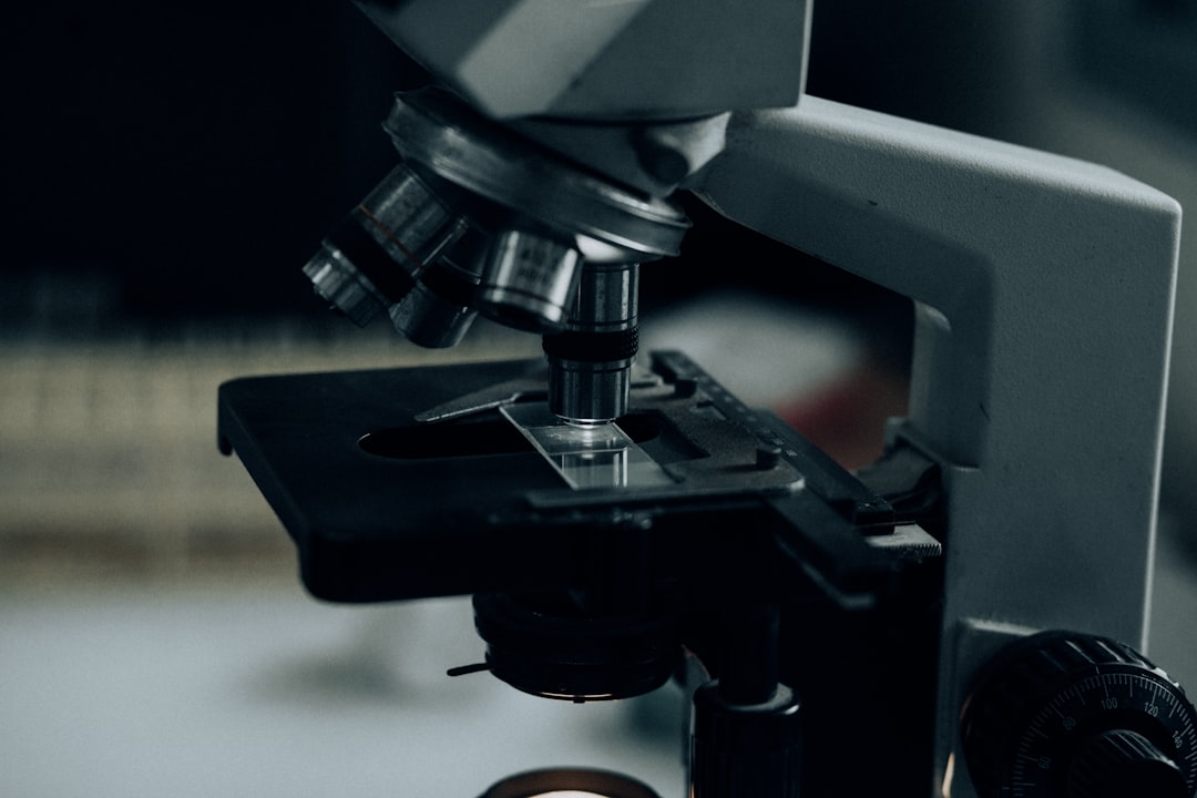
You’ve probably experienced it. That sudden, sharp sting, a surprisingly intense pain for something so seemingly innocuous. It’s the paper cut. You might be engrossed in a book, meticulously organizing files, or even just unboxing a new package. Then, unexpectedly, a thin, almost invisible edge of paper finds its way into your skin, leaving behind a trail of irritation and a surprisingly deep well of discomfort. It’s a universal experience, a testament to the silent, sharp nature of the paper we interact with daily.
This seemingly minor injury, so easily dismissed by some, can be a surprisingly complex event when you zoom far beyond the naked eye. You might think it’s a simple slice, a clean tear of the epidermis. But look closer, much closer, and you’ll discover a microscopic battlefield, a miniature drama unfolding within the layers of your skin. This isn’t just about a superficial wound; it’s about the intricate architecture of your integumentary system and the precise way a seemingly gentle material can orchestrate a surprisingly potent reaction.
You might wonder why something as mundane as paper can inflict such disproportionate pain. It’s not just about the depth of the cut, although that plays a role. It’s about how the paper’s edge interacts with the finely tuned sensory network beneath your skin. Each paper cut, therefore, becomes a unique exploration, a microscopic journey into the delicate balance between external forces and your internal biological responses.
In a fascinating exploration of the microscopic world, a related article discusses the intricate details of everyday objects when viewed under a microscope, shedding light on their hidden complexities. This article not only complements the study of paper cuts under a microscope but also expands on the broader implications of microscopic analysis in understanding material properties. For more insights, you can read the full article here: Freaky Science.
The Anatomy of Pain: Unveiling Your Skin’s Sensory Network
Before we delve into the paper’s devastating performance, let’s understand the stage upon which it plays out: your skin. It’s far more than just a barrier; it’s a dynamic, complex organ housing an incredible array of sensory receptors. This intricate network allows you to perceive the world around you, from the softest brush of a feather to the jarring impact of a fall. When that paper edge slices through, it’s not just a tear; it’s an electrical signal zapping through this finely tuned system.
The Epidermis: Your First Line of Defense
Your skin’s outermost layer, the epidermis, is your primary shield against the external world. It’s a stratified marvel, composed of several sub-layers, each with its specialized function. When you get a paper cut, it’s primarily the epidermis that’s breached.
Stratum Corneum: The Tough, Keratinized Shield
At the very top is the stratum corneum, a dense, tightly packed layer of dead skin cells rich in keratin. Think of it as a fortified wall of bricks, providing a robust defense against pathogens and dehydration. While surprisingly resilient, its flattened, scale-like cells can be separated by the sharp, albeit microscopically uneven, edge of paper. It’s the initial resistance here that contributes to that initial tearing sensation. Your nerves, however, aren’t located in this outermost, protective dead layer.
Stratum Basale: The Regenerative Foundation
Beneath the stratum corneum lies the stratum basale, the deepest layer of the epidermis. This is where the magic of regeneration happens. New skin cells are born here, constantly dividing and migrating upwards to replace the old, shedding cells from the stratum corneum. While not directly cut by the paper, the damage to the layers above sends signals to this regenerative hub, initiating the healing process.
The Dermis: Where the Real Sensation Resides
If the paper cut is deep enough to breach the epidermis, it enters the dermis, a much thicker, more vascular, and crucially, more innervated layer. This is where the true sensation of pain, itchiness, and temperature is registered.
Mechanoreceptors: Feeling the Touch
The dermis is a rich tapestry woven with various specialized nerve endings, collectively known as mechanoreceptors. These are the sensors that detect physical pressure, vibration, and stretching.
Merkel’s Discs: Fine Touch Reception
Even in the papillary dermis, just below the epidermis, you’ll find Merkel’s discs. These are slow-adapting receptors that are highly sensitive to pressure and fine touch. When the paper glides across these tiny structures, they fire signals, contributing to the immediate awareness of contact.
Meissner’s Corpuscles: Light Touch and Flutter
Deeper in the dermal papillae, you encounter Meissner’s corpuscles. These are rapidly adapting receptors, meaning they respond quickly to changes in sensation. They are responsible for detecting light touch, flutter, and are particularly adept at sensing textures. The subtle rasp of the paper edge against these can contribute to the unsettling feeling of intrusion.
Ruffini Endings: Sustained Pressure and Stretch
Further down in the reticular dermis, Ruffini endings are more attuned to sustained pressure, stretching, and even warmth. While less likely to be the primary source of pain in a superficial paper cut, if the cut has some depth, these receptors can be activated, contributing to a duller ache.
Pacinian Corpuscles: Deep Pressure and Vibration
At the deepest levels of the dermis, Pacinian corpuscles lie dormant in wait. These are large, onion-like structures that are incredibly sensitive to deep pressure and high-frequency vibrations. While typically associated with more significant impacts, they can be stimulated by the pressure pulse transmitted through the skin from the paper cut.
Nociceptors: The Pain Detectors
The most critical players in the paper cut drama are the nociceptors. These are pain receptors, a diverse group of free nerve endings that are specifically designed to detect harmful stimuli. They are found throughout the epidermis and dermis.
A-delta Fibers: Sharp, Prickly Pain
The initial, sharp, burning pain you feel from a paper cut is primarily transmitted by A-delta fibers. These are thinly myelinated nerve fibers that conduct signals relatively quickly, allowing for immediate awareness of the injury. They are responsible for that immediate, localized sting.
C-Fibers: Dull, Aching Pain
As the initial sharp pain subsides, you might experience a duller, throbbing ache. This sensation is carried by C-fibers, which are unmyelinated and conduct signals much more slowly. These fibers are crucial for signaling inflammatory responses and can contribute to the lingering discomfort of a paper cut. They are also responsible for the more diffuse, burning sensation that can follow.
The Junctional Zone: Where Epidermis Meets Dermis
The junction between the epidermis and dermis, known as the dermal-epidermal junction, is a critical area. It’s characterized by wavy projections called dermal papillae and epidermal ridges. This complex interface increases the surface area, strengthening the bond between the two layers. A paper cut, by its very nature, often tears through these delicate interdigitations, disrupting this vital connection and exposing the highly innervated dermis.
The Paper’s Edge: A Microscopic Landscape of Destruction
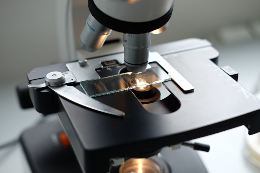
You might see a sheet of paper as a smooth, consistent surface. But at a microscopic level, even the seemingly smooth edges of paper are surprisingly irregular and rough. It’s these microscopic imperfections that turn a gentle material into a surprisingly effective cutting tool.
The Cellulose Fibers: The Building Blocks
Paper is made from cellulose fibers, which are derived from wood pulp or other plant materials. These fibers are tangled and matted together to form a cohesive sheet. During the papermaking process, these fibers are processed, sometimes bleached, and then pressed together.
The Shearing Force
When a paper edge makes contact with your skin, it’s not a clean, single slice like a scalpel. Instead, the microscopically jagged edges of the cellulose fibers act like tiny saw blades. They engage with the skin’s surface, and with even a slight lateral movement, they create a shearing force. This force pulls and tears at the skin cells.
Surface Roughness: The Unseen Sharpness
The quality of the paper’s edge plays a significant role. A freshly cut ream of paper might have a sharper, more defined edge than a well-worn book page. The manufacturing process, including the type of blade used for cutting, influences the micro-topography of the paper’s edge.
Types of Paper and Their Cutting Potential
- Cardstock and Coated Papers: These often have denser fibers and a smoother surface, but their edges can be surprisingly sharp and clean. The density can lead to a more forceful penetration.
- Recycled Papers: These might have shorter, more jagged fibers, which can create a more aggressive tearing action.
- Aged or Damaged Paper: Frayed edges, created by handling or time, can present a multitude of tiny, sharp points that can snag and lacerate the skin.
Microscopic Imperfections: The Devil is in the Details
Even a seemingly “smooth” paper edge will have microscopic peaks and valleys. These tiny irregularities, invisible to the naked eye, are precisely what interact with your skin. When the paper glides across, these points catch and tear at the Stratum Corneum, and if sufficient force is applied, they can dig into the more sensitive dermis. You can imagine it like dragging a miniature, uneven saw across your skin.
The Inflammatory Cascade: Your Body’s Response to Invasion

A paper cut, no matter how small, is an injury. Your body’s sophisticated immune system recognizes this breach in its defenses and immediately initiates a cascade of inflammatory responses. This is a vital process aimed at cleaning up the damaged tissue and initiating repair, but it’s also responsible for much of the discomfort associated with a paper cut.
Mast Cells: The First Responders
Upon detection of tissue damage, specialized immune cells called mast cells, residing in the dermis, spring into action. They are like tiny alarm bells, and when triggered, they release a cocktail of potent chemicals.
Histamine: The Redness and Swelling Agent
One of the primary chemicals released by mast cells is histamine. This molecule is a potent vasodilator, meaning it causes blood vessels to widen. This increased blood flow to the injured area is responsible for the redness and swelling you observe around a paper cut. It also makes the area more permeable, allowing other immune cells to reach the site.
Bradykinin: The Pain Amplifier
Another crucial chemical involved in the inflammatory response is bradykinin. It’s a peptide that sensitizes nerve endings, particularly the free nerve endings that are nociceptors. This sensitization means that even stimuli that wouldn’t normally be painful become capable of triggering pain signals. Bradykinin is a major contributor to the throbbing pain and tenderness of a paper cut.
Neutrophils and Macrophages: The Clean-up Crew
Following the initial release of chemicals, other immune cells are recruited to the site of injury.
Neutrophils: The Early Eliminators
Neutrophils are among the first white blood cells to arrive. They are phagocytic cells, meaning they engulf and digest cellular debris, bacteria, and other foreign material. In a paper cut, they help to clear away any damaged skin cells and any microscopic paper fragments that might have been introduced.
Macrophages: The Long-Term Cleaners and Signalers
Macrophages, which develop from monocytes, arrive later but are more long-lived. They also perform phagocytosis, but their role is broader. They not only clean up debris but also release signaling molecules (cytokines) that continue to orchestrate the inflammatory and healing processes. They essentially guide the repair efforts.
Prostaglandins: The Persistent Ache
The inflammatory process also involves the production of prostaglandins, which are lipid compounds. Prostaglandins further sensitize nociceptors, contributing to the dull, aching pain and increased tenderness that can accompany a paper cut. This contributes to the lingering discomfort that makes you constantly aware of that small wound.
In a fascinating exploration of everyday objects, a recent article discusses the intricate details of a paper cut when examined under a microscope. This examination reveals the complex structure of the skin and how it reacts to minor injuries, shedding light on the healing process. For those interested in the microscopic world, you can read more about this intriguing topic in the article found here. The insights gained from such studies not only enhance our understanding of human biology but also highlight the surprising intricacies of seemingly mundane experiences.
The Healing Process: Microscopic Repair and Regeneration
| Metric | Value |
|---|---|
| Length of paper cut | 0.5 mm |
| Depth of paper cut | 0.2 mm |
| Shape of paper cut | Irregular |
| Color of paper cut | White |
While the pain and inflammation might feel like the end of the story, they are merely the dramatic opening act for your body’s incredible capacity for repair. Once the initial damage is assessed and the site is cleared, a meticulous process of regeneration begins, aiming to restore the skin’s integrity.
Fibroblasts: The Scaffolding Builders
Once the inflammatory phase begins to subside, fibroblasts become the primary architects of repair. These versatile cells, abundant in the dermis, are responsible for producing collagen and other extracellular matrix components.
Collagen Synthesis: Rebuilding the Structure
Collagen is the most abundant protein in your body, providing structural support and elasticity to tissues. Fibroblasts deposit new collagen fibers at the site of the paper cut, forming a disorganized scar tissue initially. This new matrix serves as a scaffold, bridging the gap created by the cut.
Angiogenesis: Fueling the Repair
For the healing process to be successful, the damaged tissue needs a fresh supply of oxygen and nutrients. This is where angiogenesis comes into play.
New Blood Vessel Formation
The body signals for the growth of new blood vessels (capillaries) to sprout and extend into the healing area. These new vessels not only deliver essential components for repair but also help to carry away waste products. The increased vascularization contributes to the still-present redness in the healing wound.
Epithelialization: Sealing the Breach
The final phase of wound closure involves epithelialization, where the epidermis regenerates and covers the wound bed.
Keratinocyte Migration and Proliferation
Basal keratinocytes from the edges of the wound begin to divide and migrate across the newly formed collagen matrix. As they move, they proliferate, thickening the epidermal layer until the entire wound is covered. This process ultimately seals the breach, restoring your skin’s protective barrier.
Scar Formation: A Permanent Reminder
The scar tissue that forms from a paper cut, while less prominent than that from a larger injury, is still a testament to the repair process. It’s a patch of collagen that might have a slightly different texture and coloration than the surrounding skin. Over time, this scar tissue remodels, becoming more organized and less noticeable, but it’s a permanent biological record of the microscopic encounter between your skin and that paper edge.
The Paradox of Pain: Why Paper Cuts Hurt So Much
You might have a deeper cut from a kitchen knife, and while it’s a significant injury, the sharp, stinging pain of a paper cut often seems disproportionate. This paradox stems from a combination of factors, primarily related to your skin’s anatomy and the way the paper cuts it.
High Density of Nerve Endings in Fingertips
Your fingertips, the most common site for paper cuts, are incredibly rich in sensory receptors, especially nociceptors. This high density allows for exquisite touch discrimination, essential for manipulating objects, but it also means that even a small injury can activate a multitude of pain signals.
The “Sawing” Action
The microscopic jaggedness of the paper’s edge, as we’ve discussed, doesn’t create a clean incision. Instead, it acts like a miniature saw, dragging and tearing. This tearing motion is more disruptive to the delicate nerve endings than a clean slice, sending a more insistent and irritating signal to your brain.
Surface Area of the Cut
While a paper cut might appear very superficial, its long, thin nature can create a surprisingly large surface area of injured tissue relative to its depth. This expanded area of damaged nerve endings contributes to the overall intensity of the pain. Think of it as spreading the damage thinly over a wider area, maximizing the number of pain receptors activated.
Lubrication and Lack of Bleeding
Unlike a deeper cut where blood can lubricate the wound and wash away irritants, paper cuts are often too shallow to bleed significantly. This means the irritating paper fibers and the chemicals released by damaged cells remain in close contact with the exposed nerve endings, prolonging the sensation of pain and burning.
Infection Risk and Protective Behavior
The protective behavior your body exhibits in response to pain is essential for survival. The sharp, stinging pain of a paper cut serves as an immediate warning to withdraw your hand from the offending object. It’s a primal reflex that encourages you to protect yourself from further harm. Even the low-grade ache that follows serves as a constant reminder to avoid stressing the injured area, thus promoting healing. While the risk of infection from a paper cut is generally low, the inflammatory response is still initiated to combat any potential microbial invaders, and this inflammation itself contributes to the discomfort.
So, the next time you wince from a paper cut, remember the intricate microscopic drama that just unfolded. It’s a testament to your body’s marvelous sensory network, the surprising sharpness of everyday materials, and your remarkable capacity for healing. It’s a tiny reminder of the complex biological world that lies beneath your skin, a world that reacts to even the most seemingly insignificant of intruders.

Why Paper Cuts Hurt (It’s Not What You Think)
FAQs
What is a paper cut?
A paper cut is a small, shallow cut on the skin caused by a sharp edge of paper. It is a common and minor injury that can be quite painful due to the high number of nerve endings in the fingers.
How does a paper cut look under a microscope?
When viewed under a microscope, a paper cut appears as a jagged, irregular tear in the skin. The edges of the cut may appear frayed, and the surrounding skin may show signs of inflammation and damage.
What happens to the skin cells during a paper cut?
During a paper cut, the sharp edge of the paper tears through the layers of the skin, causing damage to the skin cells. This damage triggers a series of biological responses, including inflammation and the release of healing factors to repair the injured tissue.
How can a paper cut be treated?
To treat a paper cut, it is important to clean the wound with soap and water to prevent infection. Applying an over-the-counter antibiotic ointment and covering the cut with a bandage can help protect it as it heals. In some cases, a tetanus shot may be necessary if the cut is deep or dirty.
Can a paper cut lead to complications?
While paper cuts are generally minor injuries, they can sometimes lead to complications such as infection if not properly cleaned and cared for. It is important to monitor the cut for signs of infection, such as increased redness, swelling, or pus, and seek medical attention if necessary.