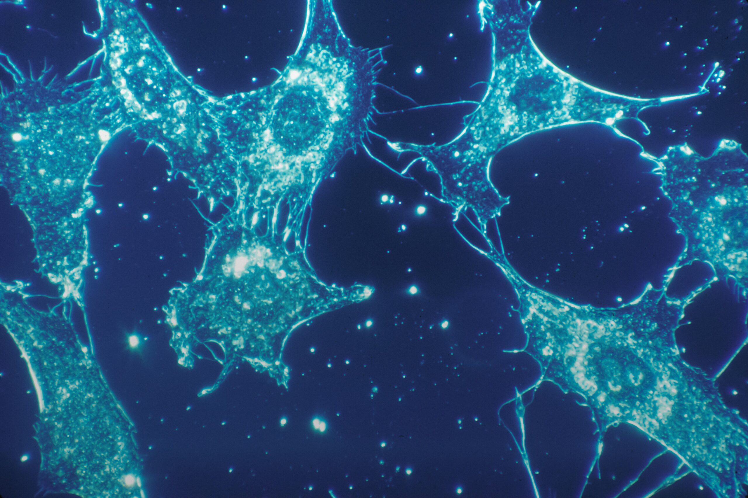
Imagine a world unseen, a microscopic universe teeming with life. This is the realm of living cells, the fundamental building blocks of all organisms. For centuries, these tiny entities remained shrouded in mystery, their inner workings invisible to the naked eye. But then, a revolution in scientific exploration dawned, one fueled by the very essence of light. You now stand at the precipice of this luminous frontier, ready to understand how light has not only illuminated the world of living cells but has also become an indispensable tool for unraveling their deepest secrets. This is the story of how living cells, once invisible, now glow with the insights brought forth by light.
Before the advent of sophisticated microscopy, your understanding of life was limited to what your eyes could perceive. The intricate dance of cells, the very engines of existence, remained hidden. It was the ingenuity of early scientists and the development of simple optical instruments that first offered a glimpse into this hidden world.
The Humble Beginnings: Early Microscopy and the Discovery of Cells
Think back to the 17th century. Imagine Robert Hooke, peering through his rudimentary microscope. His observation of cork, revealing its honeycomb-like structure, marked a pivotal moment. He coined the term “cell,” though his observations were of dead plant tissue. This initial glimpse was like a single candle flickering in a vast darkness. It sparked curiosity but offered little detail. You were only beginning to scratch the surface of what lay beneath.
Then came Antonie van Leeuwenhoek, a draper with an insatiable fascination for magnification. His meticulously crafted lenses, far superior to those of his contemporaries, allowed him to observe a menagerie of microscopic life. He saw “animalcules” in pond water, in scrapings from his teeth, and in his own blood. You can almost feel the wonder he must have experienced, encountering these bustling, independent entities for the first time. This was not just seeing, but witnessing life in its most fundamental form.
The Limitations of Early Light: Resolution and Contrast
While these early microscopes were revolutionary, they were inherently limited. The resolution – the ability to distinguish between two closely spaced points – was poor. You could see shapes, but the fine details of cellular structures remained elusive. Furthermore, most biological specimens are naturally transparent, making it difficult to discern them against their background. Imagine trying to see a delicate spiderweb against a white wall; without any contrast, it would be nearly invisible. This lack of contrast was a significant hurdle in understanding cellular morphology and function. You were looking, but you were not truly seeing the intricate machinery within.
Recent research has uncovered fascinating insights into the phenomenon of bioluminescence, revealing that living cells emit light as a byproduct of various biochemical processes. This intriguing topic is explored in detail in the article found at Freaky Science, which discusses the implications of cellular light emission for understanding biological functions and potential applications in medicine and technology.
Harnessing Light for Clarity: Advancements in Light Microscopy
The limitations of early microscopes spurred innovation. Scientists recognized that to truly understand living cells, they needed to manipulate light in more sophisticated ways to enhance visibility and reveal finer details. This led to the development of various microscopy techniques, each building upon the principles of light interaction with matter.
Bright-Field Microscopy: The Standard for Everyday Observation
This is perhaps the most common type of light microscopy, and you’ve likely encountered it in introductory biology courses. In bright-field microscopy, light from a source passes directly through the specimen. The specimen absorbs some of this light, creating contrast against the brightly illuminated background. Think of it as looking at a silhouette. While effective for many unstained cells and tissues, its limitations in contrast become apparent when you need to observe delicate intracellular structures or unstained living cells. You can see the general outline, but the subtle nuances are lost.
Phase Contrast Microscopy: Revealing the Invisible by Manipulating Light Waves
Here’s where you start to see a significant leap in your ability to observe living cells. Erwin Warneck developed phase contrast microscopy, a brilliant technique that transforms differences in refractive index within a cell into differences in light intensity. Living cells, with their varying densities and structures, have different refractive indices. Phase contrast microscopy manipulates the phase of light waves that pass through these different regions. This subtle manipulation, invisible to the naked eye, creates a discernible difference in brightness, making cellular structures pop into view without the need for staining. You are now able to observe the dynamic movements and subtle changes within living cells, a feat impossible with simple bright-field microscopy.
Differential Interference Contrast (DIC) Microscopy: Adding Depth and Dimension
Building upon the principles of phase contrast, DIC microscopy, also known as Nomarski microscopy, provides an even more striking sense of three-dimensionality. DIC uses a prism to split a beam of polarized light into two beams that are slightly offset. These beams then pass through adjacent points of the specimen. Upon recombination, interference occurs, amplifying subtle differences in the optical path length. The result is an image with a striking relief or pseudo-3D effect. Imagine looking at a sculpture with dramatic lighting; DIC microscopy provides a similar sense of depth and texture, allowing you to appreciate the subtle contours and structures within living cells. You can now discern the intricate network of organelles and the delicate movement of intracellular components with remarkable clarity.
Illuminating Beyond Visible Light: The Power of Fluorescence
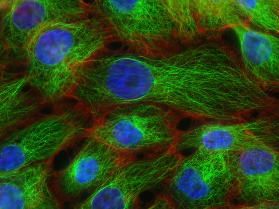
While visible light microscopy has been invaluable, many cellular processes are too subtle or occur too deep within cells to be easily visualized. This is where the magic of fluorescence microscopy truly shines. By attaching fluorescent molecules to specific cellular components, you can make them emit light of a different color when excited by a specific wavelength of light, effectively turning your cells into tiny, glowing beacons.
The Principles of Fluorescence: How Molecules Emit Light
At its core, fluorescence involves the absorption of light energy by a molecule, causing its electrons to jump to a higher energy level. As these electrons return to their ground state, they release the excess energy as light of a longer wavelength. Think of it like a glow stick: you break the inner vial, mix the chemicals, and energy is released as visible light. In fluorescence microscopy, you’re using specialized molecules, called fluorophores, and exciting them with a specific wavelength of light. The light emitted by these fluorophores reveals the location and distribution of the molecules they are attached to.
Fluorescent Proteins: Genetically Encoded Illuminators
A monumental breakthrough in fluorescence microscopy came with the discovery and engineering of fluorescent proteins, most notably Green Fluorescent Protein (GFP). You can now genetically engineer cells to produce GFP (or its variants of different colors) within specific organelles or attached to specific proteins. This means you can label virtually any component of a living cell and watch it in real-time. Imagine a cell filled with tiny, colored spotlights, each highlighting a different cellular process. This ability to tag and track specific molecules within living cells has revolutionized cell biology, allowing you to observe everything from protein trafficking to gene expression in action.
Confocal Microscopy: Zapping Away the Blur
One of the main challenges with standard fluorescence microscopy is out-of-focus light, which can blur the image and obscure fine details. Confocal microscopy elegantly solves this problem. It uses a pinhole aperture to block out-of-focus light, allowing only light from a specific focal plane to reach the detector. By scanning a laser across the specimen and collecting light from each point, you can reconstruct a sharp, in-focus image of a thin optical section of the cell. By acquiring multiple optical sections at different depths, you can create a 3D reconstruction of the cell, offering an unprecedented view of its internal architecture. You are now able to create incredibly detailed, high-resolution images of cellular structures, peering deep into the cell without disturbing its delicate balance.
Super-Resolution Microscopy: Pushing the Boundaries of Resolution
Even confocal microscopy has its limits, dictated by the wavelength of light used. However, the development of super-resolution microscopy techniques has shattered these traditional barriers. These advanced techniques, such as STORM (Stochastic Optical Reconstruction Microscopy) and PALM (Photoactivated Localization Microscopy), allow you to achieve resolutions far beyond the diffraction limit of light. They work by exciting only a sparse subset of fluorescent molecules at a time, precisely localizing each activated molecule, and then computationally reconstructing a high-resolution image. You are effectively “outsmarting” the physics of light to see details that were previously unimaginable, revealing the molecular machinery of life with astonishing clarity.
Illuminating Function: Beyond Structure to Dynamic Processes
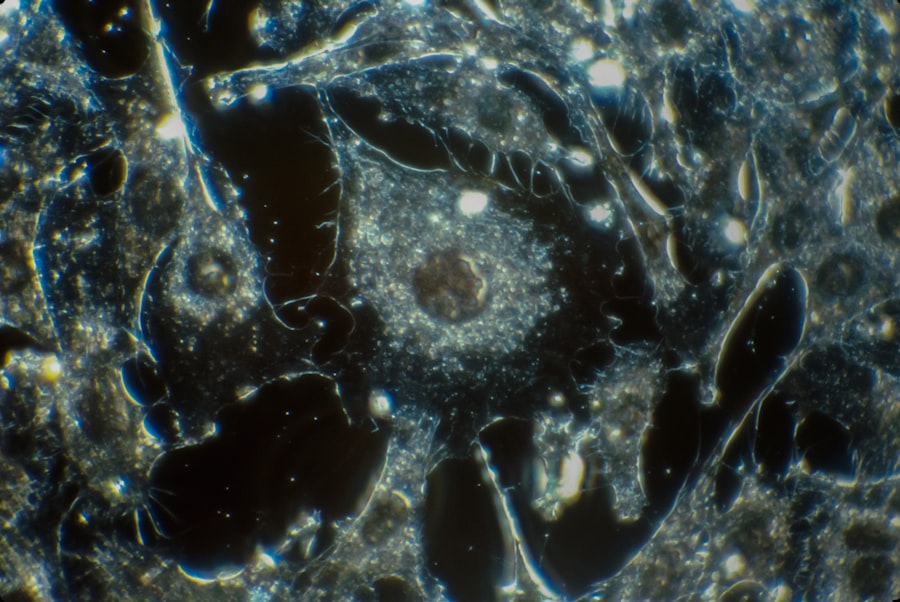
The ability to visualize cellular structures is crucial, but understanding how these structures function is the ultimate goal. Light microscopy, particularly fluorescence-based techniques, has been instrumental in illuminating the dynamic processes that occur within living cells.
Tracking Molecules in Motion: From Diffusion to Transport
Imagine a bustling city where goods are constantly being moved from one place to another. Cells are no different, with molecules constantly diffusing and being actively transported to their designated locations. Using fluorescently tagged molecules, you can observe this traffic in real-time. You can track the movement of proteins from their synthesis in the cytoplasm to their destination within organelles, or monitor the diffusion of signaling molecules across membranes. Techniques like Fluorescence Recovery After Photobleaching (FRAP) allow you to measure the rate of molecular exchange and diffusion within specific cellular compartments, giving you insights into the fluidity and dynamics of cellular components.
Visualizing Cellular Communication: Signaling Pathways in Action
Cells don’t exist in isolation; they constantly communicate with their environment and with each other. These communication pathways often involve a cascade of molecular events, each of which can be visualized using fluorescent probes. You can track the activation of signaling proteins, observe the influx of calcium ions into the cell, or monitor the binding of ligands to receptors. By labeling different components of a signaling pathway with different colored fluorescent tags, you can observe how these pathways are initiated, propagated, and ultimately lead to a cellular response. You are essentially watching the intricate language of cellular communication unfold before your eyes.
Observing Cellular Dynamics: Cell Division, Migration, and Death
The life of a cell is a journey filled with dramatic events. From the precise choreography of cell division to the purposeful movement of cells during development and disease, and the inevitable process of programmed cell death (apoptosis), these dynamic processes are now observable thanks to advanced light microscopy. You can watch as a cell replicates its DNA, divides into two daughter cells, or witness the directed migration of cells to repair damaged tissue. You can even observe the intricate molecular mechanisms that trigger apoptosis, a vital process for maintaining tissue health. These dynamic visualizations provide a profound understanding of how life itself unfolds at the cellular level.
Recent studies have revealed that living cells emit light, a phenomenon known as bioluminescence, which has intrigued scientists for years. This intriguing aspect of cellular biology not only sheds light on the inner workings of life but also opens up new avenues for research in fields such as medicine and environmental science. For a deeper understanding of this fascinating topic, you can explore a related article that discusses the implications of cellular light emission in greater detail. Check it out here: Freaky Science.
Illuminating the Path to Discovery: The Future of Light and Living Cells
| Living Cells Emit Light | Metrics |
|---|---|
| Bioluminescence | Measurement of light emitted by living organisms |
| Luciferase activity | Quantification of enzyme activity in light production |
| Bioluminescent imaging | Visualization of light emission for biological research |
The journey of light and living cells is far from over. As technology continues to advance, the possibilities for exploration are becoming increasingly exciting. You are poised to witness even more profound discoveries, driven by the relentless pursuit of understanding life at its most fundamental level.
Advancements in Probe Design: More Specific and Sensitive Labels
The development of new and improved fluorescent probes is a continuous area of research. Scientists are creating probes that are more sensitive, emit brighter light, and can be targeted to an even wider array of cellular molecules and processes. You can expect probes that can simultaneously detect multiple cellular events, offering a more holistic view of cellular function. Furthermore, the development of genetically encoded probes that can be activated by specific stimuli or report on their environment will unlock new avenues of investigation.
Integration with Other Technologies: A Multi-Modal Approach
The future of cellular illumination lies in the integration of light microscopy with other cutting-edge technologies. Imagine combining high-resolution imaging with techniques like single-cell genomics or proteomics. This multi-modal approach will allow you to correlate structural and functional observations with the genetic and molecular makeup of individual cells. You will be able to understand not just what a cell is doing, but also why it is doing it, based on its unique molecular profile.
AI and Machine Learning: Unlocking Patterns in the Data
The sheer volume of data generated by modern microscopy techniques is immense. Artificial intelligence (AI) and machine learning are emerging as powerful tools to analyze this data, identify subtle patterns that might be missed by human observation, and automate tedious tasks like image segmentation and analysis. You can envision AI-powered microscopes that can automatically identify specific cellular events, track thousands of cells simultaneously, and even predict cellular behavior based on observed patterns. This will significantly accelerate the pace of scientific discovery.
Towards Clinical Applications: Diagnosis and Therapy
The insights gained from illuminating living cells have profound implications for human health. Understanding cellular dysfunction is the cornerstone of diagnosing and treating diseases. You are already seeing the use of fluorescent imaging in diagnostic procedures and surgical guidance. In the future, light-based techniques may lead to more targeted drug delivery systems, allowing therapies to be precisely delivered to diseased cells, minimizing side effects. Furthermore, a deeper understanding of cellular processes could pave the way for regenerative medicine, enabling you to repair damaged tissues by guiding cellular behavior with light.
Your journey into the world of living cells, illuminated by the power of light, is a testament to human curiosity and ingenuity. From the earliest, rudimentary microscopes to the sophisticated super-resolution techniques of today, light has been your constant companion, revealing the secrets of life one photon at a time. As you continue to push the boundaries of what is visible, you are not just observing life; you are actively participating in its unraveling, charting a future where understanding and manipulating the cellular world holds the key to unlocking unprecedented advancements in science, medicine, and our very comprehension of existence.

You’re Emitting Light Right Now—So Why Can’t You See It?
FAQs
What is bioluminescence?
Bioluminescence is the production and emission of light by living organisms, such as fireflies, jellyfish, and certain types of fungi and bacteria.
How do living cells emit light?
Living cells emit light through a process called bioluminescence, which involves the production of light by a chemical reaction within the cells. This reaction typically involves the enzyme luciferase and a light-emitting molecule called luciferin.
What are the potential applications of bioluminescence in research and medicine?
Bioluminescence has various applications in research and medicine, including as a tool for studying gene expression, protein interactions, and cellular processes. It is also used in medical imaging techniques, such as bioluminescence imaging, to track and monitor biological processes in living organisms.
Which organisms are known to exhibit bioluminescence?
Bioluminescence is observed in a wide range of organisms, including certain species of fish, squid, jellyfish, fireflies, fungi, and bacteria. Each of these organisms uses bioluminescence for different purposes, such as attracting mates, deterring predators, or luring prey.
What are the factors that can influence the intensity and color of bioluminescent light emitted by living cells?
The intensity and color of bioluminescent light emitted by living cells can be influenced by various factors, including the type of luciferase and luciferin involved in the reaction, the availability of oxygen, pH levels, and temperature. These factors can affect the efficiency and characteristics of the bioluminescent reaction.