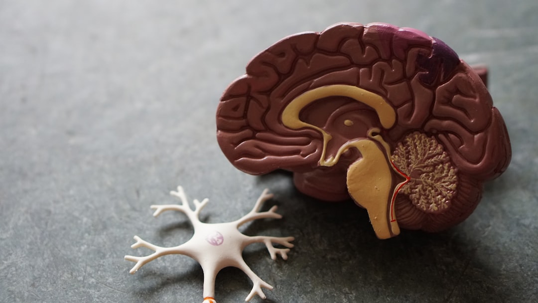
You’re probably wondering how your brain, this incredible organ ticking away inside your skull, manages to pull off all the amazing things it does. From recalling a childhood memory to deciding whether to have that second slice of pizza, it’s all thanks to a sophisticated communication system. At the heart of this system are your neurons, the fundamental building blocks of your nervous system. They’re the messengers, the electrical wires, and the chemical couriers, constantly chattering to each other to orchestrate every thought, feeling, and action you experience. Understanding how these microscopic marvels transmit signals is like peeking under the hood of your very existence.
Imagine a tiny, specialized cell, designed for one primary purpose: sending and receiving information. That’s essentially what a neuron is. It’s a far cry from your average skin cell or muscle cell; it’s a master of communication. While they come in various shapes and sizes, most neurons share a common architecture, meticulously crafted for efficient signal transmission. You’ll find billions of these cells in your brain and throughout your entire nervous system, forming an intricate network that’s responsible for everything you are.
The Three Key Players: Structure and Function
To grasp how neurons communicate, you need to understand their core components. Think of it as learning the alphabet before you can read a book.
The Cell Body (Soma): The Neuron’s Control Center
This is the main part of the neuron, containing the nucleus, which houses the cell’s genetic material, and other essential organelles like mitochondria. The cell body acts as the neuron’s metabolic powerhouse, responsible for keeping the cell alive and functioning. It also integrates all the incoming signals from other neurons. If the sum of these signals reaches a certain threshold, the cell body will initiate its own signal to be sent out.
The Dendrites: The Receiver Antennas
Branching out from the cell body like the branches of a tree are the dendrites. Their primary role is to receive signals from other neurons. Each dendrite is covered in tiny spines, further increasing their surface area for maximum signal reception. Think of them as your neuron’s listening posts, constantly on the lookout for incoming messages. The more dendrites a neuron has, the more connections it can make and the more information it can potentially receive.
The Axon: The Signal Transmitter Cable
Projecting from the cell body is a long, slender projection called the axon. This is the neuron’s communication highway, responsible for transmitting signals away from the cell body to other neurons, muscles, or glands. Axons can vary greatly in length, from mere millimeters to over a meter long, reaching across vast distances within your body.
Neurons are fascinating cells that communicate through complex signaling mechanisms, allowing for the transmission of information throughout the nervous system. For a deeper understanding of how neurons send signals, you can explore an insightful article that delves into the intricacies of neuronal communication and the role of neurotransmitters. To read more about this topic, visit this article.
The Electrical Pulse: Firing an Action Potential
The magic of neural communication lies in its combination of electrical and chemical processes. The initial spark that ignites the signal within a neuron is electrical in nature, a rapid change in electrical potential across its membrane. This electrical impulse is known as an “action potential.”
Resting Potential: The Neuron’s Neutral State
Before a neuron can fire an action potential, it needs to be in a state of readiness, much like a coiled spring. This is called the resting potential. At rest, the inside of the neuron is more negatively charged than the outside. This difference in charge is maintained by the distribution of ions (charged particles) across the neuron’s cell membrane. Specifically, there’s a higher concentration of sodium ions (Na+) outside the cell and a higher concentration of potassium ions (K+) inside the cell. This uneven distribution is actively maintained by specialized protein pumps embedded in the membrane, which require energy to operate. This creates a crucial electrochemical gradient that the neuron can harness.
Depolarization and Repolarization: The Electrical Wave
When a neuron receives a sufficient excitatory signal from another neuron, it triggers a cascade of events.
The Influx of Sodium Ions: The Rising Phase
Excitatory signals cause ion channels in the neuron’s membrane to open, allowing positively charged sodium ions (Na+) to rush into the cell down their concentration gradient. This influx of positive charge makes the inside of the neuron less negative, a process called depolarization. As more sodium channels open and more sodium ions enter, the membrane potential rapidly becomes positive. This rapid reversal of charge is the initial surge of the action potential.
The Efflux of Potassium Ions: The Falling Phase
Immediately following the influx of sodium, potassium channels open, allowing positively charged potassium ions (K+) to flow out of the cell. This efflux of positive charge makes the inside of the neuron negative again, a process called repolarization. It’s like the electrical charge is being discharged, resetting the neuron for its next potential firing.
Hyperpolarization: The Undershoot
Sometimes, the potassium channels remain open a little longer, causing a brief period where the inside of the neuron becomes even more negative than its resting potential. This is called hyperpolarization or the undershoot. This phase is important because it makes it harder for the neuron to fire another action potential immediately, ensuring that the signal travels in one direction.
The All-or-None Principle: No Half-Measures
A crucial concept to grasp about action potentials is the “all-or-none” principle. This means that once the stimulus reaches a certain threshold, the action potential will fire with the same amplitude and duration, regardless of the strength of the stimulus. There’s no “stronger” or “weaker” action potential. It either happens fully, or it doesn’t happen at all. Think of a light switch; it’s either on or off, not somewhere in between.
The Myelin Sheath: Insulating the Fast Lane

While action potentials are fast, some axons are particularly long, and rapid transmission is crucial for many of our bodily functions. To speed up this electrical signal, many axons are wrapped in a fatty insulating layer called the myelin sheath. This sheath isn’t continuous; it’s interrupted at regular intervals by gaps called the nodes of Ranvier.
The Role of Glial Cells: Building the Sheath
The myelin sheath is actually formed by specialized glial cells. In the central nervous system (brain and spinal cord), these are called oligodendrocytes, and in the peripheral nervous system, they are Schwann cells. These cells wrap their membranes around the axon multiple times, creating layers of myelin that act as an electrical insulator.
Saltatory Conduction: Leaping Across the Gaps
The myelin sheath, with its interruptions at the nodes of Ranvier, plays a critical role in speeding up signal transmission through a process called saltatory conduction. Instead of the action potential traveling smoothly down the entire length of the axon, it “jumps” from one node of Ranvier to the next. The electrical current flows within the axon, and the insulating myelin sheath prevents the charge from leaking out. When the charge reaches a node of Ranvier, it’s strong enough to trigger the opening of ion channels there, reinitiating the action potential. This jumping process is significantly faster than continuous propagation along an unmyelinated axon.
The Synapse: Where Neurons Meet and Greet
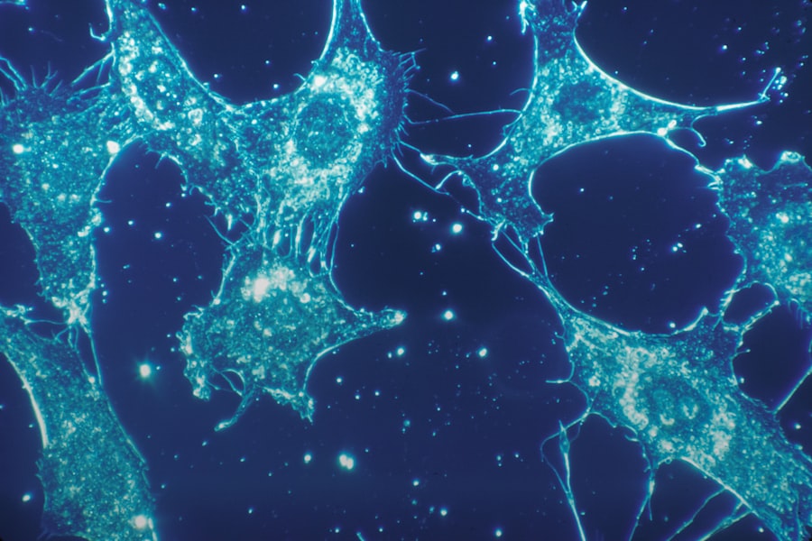
So, you’ve got this electrical signal zipping down the axon. But how does it get from one neuron to the next? This is where the synapse comes in. The synapse is the junction between two neurons, or between a neuron and an effector cell (like a muscle or gland). It’s a tiny gap, a crucial interface where communication occurs, and it’s where the electrical signal transforms into a chemical one.
The Presynaptic and Postsynaptic Neurons: The Two Sides of the Gap
At the synapse, you have two main players: the presynaptic neuron and the postsynaptic neuron.
The Presynaptic Terminal: The Signal Sender’s End
The end of the axon of the presynaptic neuron branches out into small bulbs called synaptic terminals or axon terminals. These terminals contain tiny sacs called synaptic vesicles, which are filled with chemical messengers known as neurotransmitters.
The Synaptic Cleft: The Communication Gap
The space between the presynaptic terminal and the postsynaptic neuron is called the synaptic cleft. This is a narrow gap, typically around 20-50 nanometers wide, that the neurotransmitters must cross to reach their destination.
The Postsynaptic Membrane: The Signal Receiver’s Surface
On the other side of the synaptic cleft is the membrane of the postsynaptic neuron, or the dendrite or cell body it connects to. This membrane is studded with specialized receptor proteins that are designed to bind with specific neurotransmitters.
Neurons communicate through a complex process of sending signals that involves both electrical impulses and chemical neurotransmitters. This intricate signaling mechanism allows for the rapid transmission of information throughout the nervous system, playing a crucial role in everything from reflexes to complex thought processes. For a deeper understanding of how these signals are transmitted and the underlying science, you can read more in this informative article on Freaky Science.
Chemical Messengers: The Neurotransmitters
| Aspect | Details |
|---|---|
| Neuron Structure | Neurons have a cell body, dendrites, and an axon. |
| Synapse | Neurons communicate at synapses, where neurotransmitters are released. |
| Action Potential | Neurons transmit signals through action potentials, which are electrical impulses. |
| Neurotransmitters | Chemical messengers that transmit signals across synapses. |
| Receptor Activation | Neurotransmitters bind to receptors on the receiving neuron, leading to signal transmission. |
When an action potential reaches the synaptic terminal of the presynaptic neuron, it triggers the release of neurotransmitters into the synaptic cleft. These chemical molecules are the key to crossing the gap and relaying the message to the next neuron.
Vesicle Fusion and Neurotransmitter Release: The Chemical Exchange
The arrival of the electrical signal (action potential) at the presynaptic terminal causes voltage-gated calcium channels to open. Calcium ions (Ca2+) then rush into the terminal. This influx of calcium triggers a cascade of events that leads to the fusion of synaptic vesicles with the presynaptic membrane. As the vesicles fuse, they release their neurotransmitter contents into the synaptic cleft.
Binding to Receptors: The Lock and Key Mechanism
Once in the synaptic cleft, neurotransmitter molecules drift across and bind to their specific receptor proteins on the postsynaptic membrane. This binding is often described as a lock-and-key mechanism, where a specific neurotransmitter fits precisely into its corresponding receptor. This binding event is what alters the electrical state of the postsynaptic neuron.
Excitatory vs. Inhibitory Signals: Steering the Course
The effect of the neurotransmitter binding depends on the type of receptor it binds to and the neurotransmitter itself.
Excitatory Neurotransmitters: Firing Up the Next Neuron
Some neurotransmitters, when they bind to their receptors, cause the opening of ion channels that allow positively charged ions (like sodium) to enter the postsynaptic neuron. This influx of positive charge depolarizes the postsynaptic membrane, making it more likely to fire its own action potential. These are called excitatory neurotransmitters, and they essentially encourage the next neuron to communicate. Examples include glutamate and acetylcholine.
Inhibitory Neurotransmitters: Calming Down the Chatter
Other neurotransmitters, when they bind to their receptors, cause the opening of ion channels that allow negatively charged ions (like chloride) to enter the postsynaptic neuron, or positively charged ions (like potassium) to leave. This influx of negative charge or efflux of positive charge hyperpolarizes the postsynaptic membrane, making it less likely to fire an action potential. These are called inhibitory neurotransmitters, and they essentially dampen the activity of the next neuron. Examples include GABA (gamma-aminobutyric acid) and glycine.
Neurotransmitter Clearance: Resetting the System
Once the neurotransmitters have done their job, they need to be cleared from the synaptic cleft to prevent continuous stimulation or inhibition of the postsynaptic neuron. This clearance can happen in a few ways:
Enzymatic Degradation: Breaking Down the Message
Some neurotransmitters are broken down by specific enzymes present in the synaptic cleft. For example, acetylcholine is degraded by the enzyme acetylcholinesterase.
Reuptake: Recycling the Messengers
Many neurotransmitters are reabsorbed back into the presynaptic terminal by specialized transporter proteins. This process is called reuptake, and it allows the presynaptic neuron to recycle and reuse the neurotransmitters.
Diffusion: Fading Away
Some neurotransmitters simply diffuse away from the synaptic cleft into the surrounding tissue, where they are gradually metabolized.
This intricate process of neurotransmitter release, binding, and clearance ensures that neural communication is precise, timed, and under control. It’s a dynamic system that allows your brain to process information with incredible speed and complexity. You can see how this system is fundamental to every single aspect of your existence, from the simple act of breathing to the most profound emotions and complex cognitive functions.

Why Paper Cuts Hurt (It’s Not What You Think)
FAQs
What are neurons?
Neurons are specialized cells in the nervous system that transmit information through electrical and chemical signals. They are the basic building blocks of the nervous system and play a crucial role in communication within the body.
How do neurons send signals?
Neurons send signals through a process called neurotransmission. When an electrical signal, known as an action potential, reaches the end of a neuron (the axon terminal), it triggers the release of chemical messengers called neurotransmitters. These neurotransmitters then travel across the synapse to the next neuron, where they bind to specific receptors and transmit the signal.
What is the role of neurotransmitters in signal transmission?
Neurotransmitters are chemical messengers that play a key role in signal transmission between neurons. They are released from the axon terminal of one neuron and bind to receptors on the dendrites or cell body of the next neuron, allowing the signal to be transmitted from one neuron to another.
What are some common neurotransmitters?
Some common neurotransmitters include dopamine, serotonin, acetylcholine, and gamma-aminobutyric acid (GABA). Each neurotransmitter has specific functions and plays a role in various physiological processes, including mood regulation, muscle movement, and memory.
What happens when there is a disruption in neurotransmission?
Disruptions in neurotransmission can lead to various neurological and psychiatric disorders, such as Parkinson’s disease, depression, and schizophrenia. These disruptions can be caused by factors such as genetic mutations, drug abuse, or neurodegenerative diseases, and can have significant impacts on an individual’s overall health and well-being.