The human eye, a marvel of biological engineering, possesses an astonishing ability to perceive the world around us, even under challenging lighting conditions. While our vision is not as acute as that of nocturnal predators, humans have developed a sophisticated system for navigating and functioning in low light. This ability, often referred to as “night vision,” is a complex interplay of specialized cells, chemical reactions, and neural processing. Understanding how this remarkable faculty works unveils the intricate science behind seeing in the dark.
The journey of light into sight begins with the eye’s intricate structure. This organ, roughly spherical, is designed to capture light, focus it, and convert it into electrical signals that the brain can interpret. The path of light is a carefully orchestrated process, starting from the outside and moving inward.
The Cornea: The Eye’s First Lens
Light first encounters the cornea, the transparent outer layer at the front of the eye. The cornea’s curved surface acts as the eye’s primary refractive surface, bending incoming light rays. This initial bending is crucial for focusing the light towards the internal structures of the eye. Despite its critical role in vision, the cornea is avascular, meaning it has no blood vessels, receiving its nutrients from tears and the aqueous humor behind it.
The Iris and Pupil: Controlling Light Entry
Following the cornea, light passes through the pupil, the adjustable opening in the center of the iris. The iris, the colored part of the eye, functions like the diaphragm of a camera, controlling the size of the pupil. In bright light, the iris constricts the pupil, limiting the amount of light entering the eye to prevent overstimulation and damage to the retina. Conversely, in dim light, the iris dilates the pupil, allowing more light to enter, thus maximizing the visual information available to the retina. This dynamic regulation is a fundamental aspect of our visual system’s adaptability.
The Lens: Fine-Tuning Focus
Behind the iris and pupil lies the lens, a transparent, flexible structure. The lens fine-tunes the focus of light onto the retina. Unlike the cornea, which provides fixed refraction, the lens can change its shape through a process called accommodation. Ciliary muscles surrounding the lens contract or relax, altering its curvature and adjusting the focal point for objects at varying distances. While this is vital for sharp vision at all distances, it also plays a role in how effectively light is concentrated on the retina in low-light conditions.
The Retina: The Light-Sensitive Canvas
The final destination for incoming light is the retina, a thin layer of tissue lining the back of the eye. This is where the magic of converting light into neural signals truly happens. The retina is a complex neural network composed of several layers of specialized cells, including photoreceptors, bipolar cells, and ganglion cells. The photoreceptor cells, rods and cones, are the primary sensors of light. The retina’s structure and the distribution of these cells are key to understanding how we see in both bright and dim light.
Human night vision is a fascinating topic that delves into the complex mechanisms our eyes employ to perceive low-light environments. For a deeper understanding of this subject, you can explore a related article that discusses the science behind night vision and how our eyes adapt to darkness. To read more, visit this article.
Photoreceptors: The Eyes’ Light Detectors
The retina’s ability to detect light hinges on its two main types of photoreceptor cells: rods and cones. These cells contain light-sensitive pigments that undergo chemical changes when exposed to photons of light, initiating the visual signal. Their distinct properties and distribution within the retina dictate the characteristics of our vision under different lighting conditions.
Rods: Masters of Low Light
Rods are the workhorses of our night vision. They are far more numerous than cones, with approximately 120 million rods in each human eye, primarily concentrated in the peripheral regions of the retina. Rods are incredibly sensitive to light, capable of detecting even a single photon. This extreme sensitivity is due to their photopigment, rhodopsin, which is highly efficient at capturing light energy. However, rods have a significant limitation: they do not detect color. When we are in very dim light, our vision shifts to shades of gray, a testament to the dominance of rod activity.
Rhodopsin: The Key to Scotopic Vision
Rhodopsin is a complex molecule consisting of a protein called opsin and a light-absorbing molecule called retinal. Retinal is derived from vitamin A. When light strikes rhodopsin, it causes a conformational change in the retinal molecule, a process called isomerization. This change triggers a cascade of biochemical events within the rod cell, ultimately leading to the generation of an electrical signal. This signal is then transmitted to other neurons in the retina and eventually to the brain. The regeneration of rhodopsin is a relatively slow process, meaning that after exposure to bright light, rods take time to become fully functional again in dim conditions – a phenomenon known as dark adaptation.
The Scotopic Vision System
The visual system that operates primarily through rods is known as scotopic vision. This system is characterized by high sensitivity but low spatial resolution and a lack of color perception. While scotopic vision allows us to detect movement and general shapes in darkness, it is not sufficient for detailed tasks like reading or distinguishing fine features. The high convergence of signals from many rods onto a single bipolar cell contributes to this low resolution, as it amplifies the signal but blurs the detail.
Cones: Champions of Color and Detail
Cones, on the other hand, are responsible for our color vision and sharp, detailed sight. They are less numerous than rods, with about 6 million cones per eye, concentrated in the fovea, the central part of the retina. There are three types of cones, each containing a different opsin pigment that is most sensitive to specific wavelengths of light: red, green, and blue. By comparing the signals from these different cone types, the brain perceives a vast spectrum of colors.
Photopsins and Trichromatic Color Vision
The light-sensitive pigments in cones are called photopsins. Each of the three cone types possesses a specific photopsin that absorbs light most effectively in the short (blue), medium (green), or long (red) wavelengths of the visible spectrum. When light of a particular wavelength hits the retina, it stimulates these cones to varying degrees. The brain then interprets these differential stimulation patterns as specific colors. This system is known as trichromatic color vision because it relies on the three primary color-sensing mechanisms.
The Photopic Vision System
The visual system that operates primarily through cones is called photopic vision. This system is characterized by high acuity (sharpness of vision), excellent color perception, and good detail discrimination. However, cones are much less sensitive to light than rods, requiring brighter conditions to function effectively. This is why, in very dim light, our color vision disappears, and we rely almost entirely on our rods.
The Process of Dark Adaptation: Becoming Acclimated to the Dark

The transition from bright light to darkness is not instantaneous for our vision. Instead, it involves a gradual process of adaptation, where the eye adjusts its sensitivity to the diminished light levels. This adaptation, known as dark adaptation, is crucial for maximizing our ability to see in low-light environments.
The Initial Flicker: Cone Fatigue and Rod Engagement
Upon entering a dark environment from a brightly lit one, our initial vision is poor. The cones, which were highly stimulated in the light, become fatigued, and their signals diminish. Simultaneously, the rhodopsin in the rods, which was bleached by bright light, begins to regenerate. For a short period, our vision is largely reliant on the less-sensitive cones, resulting in a dim and blurry perception.
The Gradual Improvement: Rhodopsin Regeneration and Sensitivity Increase
As time passes in the darkness, the rods begin to regain their sensitivity. The regeneration of rhodopsin is the primary driver of this improvement. With more rhodopsin available, the rods become increasingly capable of detecting the faint light present. This process can take anywhere from 20 to 45 minutes to reach its full potential, during which our ability to see in the dark steadily improves. The initial phase of dark adaptation, dominated by cone recovery and a slight increase in rod sensitivity, is relatively quick, while the slower, more significant increase in sensitivity is due to rhodopsin regeneration.
Factors Influencing Dark Adaptation
Several factors can influence the speed and effectiveness of dark adaptation. Age, for instance, can play a role, with older individuals sometimes experiencing a slower adaptation process. Nutritional status is also important, particularly vitamin A intake, as it is a crucial component of rhodopsin. Exposure to certain medications or medical conditions can also impact dark adaptation. Furthermore, the intensity and duration of prior light exposure can affect how quickly the rods become functional again.
Neural Pathways: Transmitting the Visual Signal to the Brain
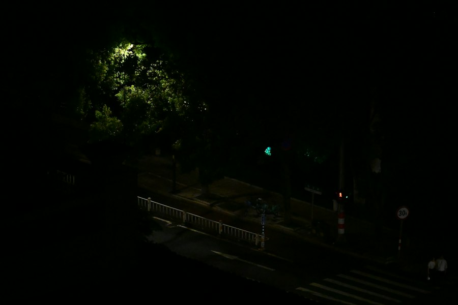
Once light has been converted into electrical signals by the photoreceptors in the retina, these signals embark on a journey through a complex network of neurons before reaching the brain for interpretation. This intricate neural processing is essential for constructing our visual perception.
From Photoreceptors to Bipolar Cells
The electrical signals generated by rods and cones are passed on to the next layer of neurons in the retina: the bipolar cells. These cells act as intermediaries, integrating the signals from multiple photoreceptors. There are different types of bipolar cells, some receiving input from rods and others from cones, further processing the visual information.
The Role of Ganglion Cells and the Optic Nerve
Bipolar cells, in turn, transmit their signals to ganglion cells. The axons of the ganglion cells converge to form the optic nerve, a thick bundle of nerve fibers that exits the back of the eye. Each optic nerve carries visual information from one eye to the brain. The optic nerve is essentially a highway for visual data, conveying the processed electrical impulses to higher visual centers.
Visual Processing in the Brain: From Signal to Perception
The optic nerve fibers travel to the brain, primarily to a region called the lateral geniculate nucleus (LGN) in the thalamus. From the LGN, the visual information is relayed to the primary visual cortex (V1) located in the occipital lobe at the back of the brain. Here, the raw electrical signals begin to be interpreted and reconstructed into meaningful images. Further processing occurs in a hierarchy of visual areas, allowing us to recognize objects, perceive depth, and understand our surroundings. This sophisticated neural circuitry is responsible for our conscious perception of sight.
Human night vision is a fascinating topic that explores how our eyes adapt to low-light conditions, allowing us to see in the dark. This ability is primarily due to the presence of rod cells in the retina, which are more sensitive to light than cone cells. For those interested in delving deeper into the science behind our vision, you can check out a related article on this subject at Freaky Science, where you can learn more about the mechanisms that enable us to navigate our surroundings when the sun goes down.
Limits of Human Night Vision: Why We Aren’t Owls
| Aspect | Description |
|---|---|
| Photoreceptors | Specialized cells in the retina called rods and cones are responsible for detecting light and transmitting signals to the brain. |
| Adaptation | Human eyes can adapt to low light conditions through a process called dark adaptation, where the sensitivity of the rods increases. |
| Peripheral vision | While rods are responsible for peripheral vision and low-light vision, cones are responsible for color vision and central vision. |
| Visual pigment | Rhodopsin, a visual pigment in the rods, is responsible for the initial detection of light in low-light conditions. |
| Optic nerve | Signals from the photoreceptors are transmitted to the brain through the optic nerve, where they are processed to form the visual perception of the environment. |
Despite the remarkable adaptations of our eyes, human night vision has inherent limitations. Compared to many nocturnal animals, our ability to see in very low light is relatively poor. Understanding these limitations helps to appreciate the evolutionary trade-offs and the specific design of our visual system.
Sensitivity vs. Acuity Trade-off
A fundamental trade-off in vision is between light sensitivity and visual acuity. Rods, with their high sensitivity, are excellent for detecting faint light but have poor acuity, leading to blurry vision and a lack of color. Cones, with their high acuity and color perception, require much brighter light to function. This means that when light levels drop significantly, we sacrifice detail and color for the ability to simply detect the presence of objects.
The Absence of a Tapetum Lucidum
Many nocturnal animals possess a reflective layer behind the retina called the tapetum lucidum. This layer acts like a mirror, bouncing light that has passed through the retina back onto the photoreceptors. This gives the photoreceptors a second chance to absorb photons, effectively increasing the amount of light available for vision. Humans, like most primates, lack a tapetum lucidum. This absence contributes to our lower sensitivity in dim light conditions, as photons that pass through the retina are not reflected back for further capture.
Color Perception in Low Light
As mentioned earlier, our color vision is significantly diminished, if not entirely absent, in very low light conditions. This is because cones, responsible for color perception, are much less sensitive than rods. In dim light, our visual system relies almost exclusively on rods, which are monochromatic. This shift from trichromatic to monochromatic vision is a key limitation of human night vision.
Peripheral vs. Central Vision in the Dark
Due to the distribution of rods and cones, our peripheral vision is actually better in dim light than our central (foveal) vision. The high concentration of rods in the peripheral retina allows us to detect movement and faint shapes in the shadows. However, the fovea, which is densely packed with cones and responsible for sharp, detailed vision, becomes largely non-functional in darkness. This explains why we often notice movement in our peripheral vision in the dark, but struggle to identify what it is when we try to look at it directly.
In conclusion, human night vision is a testament to the intricate design and adaptability of our visual system. From the light-gathering capabilities of our eyes to the complex neural processing in the brain, every component plays a crucial role in enabling us to perceive our world, even when the lights are low. While we may not possess the supernatural vision of nocturnal creatures, our ability to see in the dark, albeit in shades of gray and with reduced detail, is a remarkable feat of biological engineering. The science behind this ability continues to be a source of fascination, highlighting the elegant interplay of physics, chemistry, and biology that allows us to experience the world through sight.

The Space Photos Are Real—But Your Eyes Would See This
FAQs
1. How does human night vision work?
Human night vision works through a process called dark adaptation, where the eyes gradually become more sensitive to low light conditions. This is due to the presence of rod cells in the retina, which are responsible for detecting light and allowing us to see in dimly lit environments.
2. What role do rod cells play in human night vision?
Rod cells are highly sensitive to light and are responsible for allowing us to see in low light conditions. They contain a pigment called rhodopsin, which is able to detect even small amounts of light and send signals to the brain to create an image.
3. How long does it take for the human eye to adjust to darkness?
It takes approximately 20-30 minutes for the human eye to fully adjust to darkness. During this time, the rod cells in the retina gradually become more sensitive to low light conditions, allowing us to see more clearly in the dark.
4. What factors can affect human night vision?
Factors such as age, genetics, and overall eye health can affect human night vision. Older individuals may experience a decline in night vision due to changes in the eye’s structure, while certain genetic conditions can impact the function of rod cells. Additionally, conditions such as cataracts or vitamin deficiencies can also affect night vision.
5. Can night vision be improved through external means?
While there are devices such as night vision goggles that can enhance our ability to see in the dark, there are no proven methods to permanently improve human night vision. However, maintaining overall eye health, including regular eye exams and a balanced diet, can help support optimal night vision.