Parkinson’s disease, a neurodegenerative disorder primarily affecting motor control, presents a significant diagnostic challenge. While clinical assessment remains the cornerstone of diagnosis, it is retrospective, relying on the manifestation of motor symptoms that often appear when substantial neuronal loss has already occurred. This inherent delay has spurred a relentless pursuit for earlier, more definitive diagnostic markers. Among the burgeoning array of potential tools, the skin biopsy has emerged as a beacon of promise, offering a tangible window into the pathological processes unfolding within the nervous system. This article explores the science behind skin biopsies in Parkinson’s diagnosis, detailing their current utility, evolving applications, and the exciting future they hold.
At the heart of Parkinson’s disease pathology lies the abnormal aggregation of a protein called alpha-synuclein into Lewy bodies and Lewy neurites. These proteinaceous clumps are considered a hallmark of the disease, disrupting normal neuronal function and ultimately leading to cell death.
The Ubiquitous Nature of Alpha-Synuclein
While primarily associated with the brain, alpha-synuclein is also found in peripheral tissues, including the skin. This widespread distribution, once thought to be primarily functional in synaptic regulation, has proven to be a critical insight. The skin, in essence, acts as an accessible extension of the nervous system’s biochemical landscape.
Alpha-Synuclein in the Skin: A Microscopic Reflection
Research has demonstrated that alpha-synuclein pathology, in the form of phosphorylated alpha-synuclein and protein aggregates, can be detected in cutaneous nerves. These nerves, which innervate various structures within the skin, including sweat glands, hair follicles, and blood vessels, are susceptible to the same pathological processes that afflict neurons in the brain. Therefore, the presence and characteristics of alpha-synuclein in these cutaneous nerves can serve as a surrogate marker for the systemic involvement characteristic of Parkinson’s disease. This is akin to finding a single dropped stitch on a sweater that hints at a larger unraveling within the garment.
Differentiating Parkinson’s from Other Synucleinopathies
A key advantage of skin biopsy analysis lies in its potential to differentiate Parkinson’s disease from other neurodegenerative disorders that also involve alpha-synuclein aggregation, collectively termed synucleinopathies. Conditions such as Multiple System Atrophy (MSA) and Dementia with Lewy Bodies (DLB) share this pathological hallmark but present with distinct clinical features and prognoses. Precise differentiation is crucial for accurate patient management and the development of targeted therapies.
Recent advancements in the field of Parkinson’s disease diagnosis have highlighted the potential of skin biopsies as a non-invasive method for identifying the condition. A related article discusses how skin biopsies can reveal the presence of alpha-synuclein aggregates, which are indicative of Parkinson’s. For more information on this innovative approach, you can read the article here: Skin Biopsy for Parkinson’s Diagnosis. This method could pave the way for earlier and more accurate diagnoses, ultimately improving patient outcomes.
The Diagnostic Process: From Skin Scraping to Cellular Analysis
The journey of a skin biopsy from a patient to a diagnostic insight is a multi-step process, each stage meticulously designed to extract and analyze the relevant pathological information. While the biopsy itself is a relatively minor surgical procedure, the subsequent laboratory analysis is where the magic truly happens.
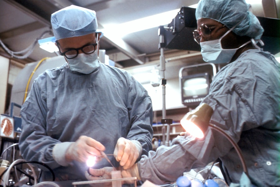
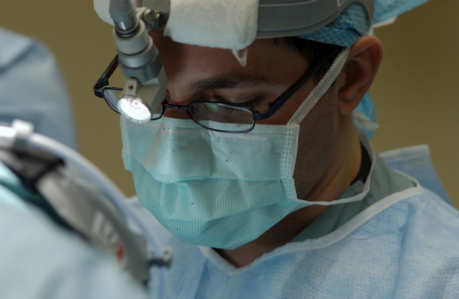
The Biopsy Procedure: A Minimal Intervention
Typically, a small sample of skin, usually a few millimeters in diameter, is obtained under local anesthesia. This can be performed from various sites, with the nape of the neck and the thigh being common locations. The procedure is generally well-tolerated and is often carried out in an outpatient setting. The brevity and relative ease of obtaining this sample make it a significantly less invasive option compared to traditional neurological investigations.
Tissue Processing: Preserving Cellular Integrity
Following collection, the skin sample undergoes meticulous processing to preserve the delicate cellular structures and the integrity of the proteins within. This often involves fixation in specific solutions to prevent degradation and ensure that the alpha-synuclein present is in a state that can be readily detected by subsequent analytical techniques.
Immunofluorescence and Immunohistochemistry: Illuminating the Pathology
The core of the diagnostic analysis relies on sophisticated staining techniques. Immunofluorescence and immunohistochemistry are two primary methods employed. These techniques utilize antibodies specifically designed to bind to alpha-synuclein. When these antibodies bind, they are tagged with fluorescent molecules or enzymes that can be visualized under a microscope.
- Immunofluorescence: This technique employs antibodies conjugated with fluorescent dyes. When excited by specific wavelengths of light, these dyes emit light of a different color, allowing researchers to pinpoint the location of alpha-synuclein within the skin tissue. Different colors can be used to highlight other cellular structures, providing crucial context.
- Immunohistochemistry: In this method, antibodies are linked to enzymes. When a suitable substrate is added, the enzyme catalyzes a reaction that produces a colored precipitate at the site of antibody binding. This colored precipitate makes the alpha-synuclein visible as a distinct stain under a light microscope.
Quantifying and Characterizing Alpha-Synuclein Deposits
Beyond simply detecting the presence of alpha-synuclein, advanced analytical techniques allow for its quantification and characterization. Researchers can assess the density of alpha-synuclein deposits, their distribution within different cutaneous nerve populations (e.g., sympathetic, sensory), and even their specific phosphorylated or oligomeric forms, which are believed to be more toxic. This level of detail provides a more nuanced understanding of the disease’s molecular underpinnings.
Clinical Applications: Current and Emerging Roles
The utility of skin biopsies in the diagnosis of Parkinson’s disease is steadily expanding, moving from a research tool to a recognized clinical application.
Supporting a Clinical Diagnosis: A Biopsy as a Confirmation
In situations where a clinical diagnosis of Parkinson’s disease is suspected but not definitively established, a positive skin biopsy revealing alpha-synuclein pathology can provide crucial corroborative evidence. This is particularly valuable in early stages of the disease when motor symptoms might be subtle or atypical. It can help to anchor the diagnosis and guide subsequent management strategies.
Differential Diagnosis: Distinguishing Synucleinopathies
As mentioned previously, the ability to identify alpha-synuclein pathology in the skin is paramount for differentiating Parkinson’s disease from other synucleinopathies. For instance, the distribution and type of alpha-synuclein deposits can differ between Parkinson’s, MSA, and DLB, offering a cellular fingerprint to distinguish these conditions. This is like having a detective with specialized tools to identify the culprit even when several suspects are present.
Identifying Prodromal Parkinson’s: A Glimpse into the Pre-Motor Phase
One of the most exciting frontiers for skin biopsy analysis is its potential to identify individuals in the prodromal stage of Parkinson’s disease, before the onset of overt motor symptoms. Non-motor symptoms, such as REM sleep behavior disorder, anosmia (loss of smell), and constipation, can precede motor manifestations by years, or even decades. Research suggests that alpha-synuclein pathology may already be present in the peripheral nervous system during this pre-motor phase.
- REM Sleep Behavior Disorder (RBD): Individuals with RBD, a condition characterized by the acting out of dreams during sleep, are at a significantly increased risk of developing Parkinson’s disease. Detecting alpha-synuclein in cutaneous nerves of individuals with RBD can serve as an early warning sign.
- Anosmia: A diminished or lost sense of smell is another common early symptom of Parkinson’s. Skin biopsies of olfactory nerve endings or related structures could potentially provide a diagnostic marker in these individuals.
Identifying individuals at high risk during these prodromal stages opens up unprecedented opportunities for early intervention and the development of neuroprotective therapies aimed at slowing or halting disease progression before significant neuronal damage occurs. This is like spotting a tiny crack in a dam before it bursts, allowing for timely repairs.
Limitations and Challenges: Navigating the Road Ahead
Despite its considerable promise, the widespread clinical adoption of skin biopsies for Parkinson’s diagnosis is not without its hurdles. Addressing these limitations is crucial for realizing its full potential.
Standardization of Protocols: Ensuring Consistency
One of the key challenges is the lack of fully standardized protocols for tissue collection, processing, and analysis across different research centers and laboratories. Variations in these procedures can lead to differences in results, impacting the reliability and reproducibility of the diagnostic findings. Establishing universally accepted guidelines is essential for widespread clinical implementation.
Interpretation of Results: Subjectivity and Expertise
While objective markers are sought, the interpretation of alpha-synuclein staining can sometimes involve a degree of subjective assessment by expert neuropathologists. Subtle differences in the pattern, intensity, or location of deposits might require significant expertise to interpret accurately. Developing quantitative and automated analysis methods can help to mitigate this.
False Positives and False Negatives: The Nuances of Diagnosis
Like any diagnostic test, skin biopsies are susceptible to obtaining false positive or false negative results. Factors such as the specific site of biopsy, the age of the individual, and the presence of other skin conditions could potentially influence the findings. Further research is needed to fully understand the sensitivity and specificity of skin biopsies in various clinical scenarios. The absence of definitive Parkinson’s pathology in a skin biopsy does not entirely rule out the disease, and vice versa.
Cost-Effectiveness and Accessibility: Bridging the Gap
The cost associated with laboratory analysis, particularly for advanced techniques like immunohistochemistry, can be a barrier to widespread accessibility. Ensuring that this diagnostic tool is cost-effective and readily available to a broad patient population, especially in resource-limited settings, is a critical consideration for its future integration into routine clinical practice.
Recent advancements in the field of neurology have highlighted the potential of skin biopsies as a diagnostic tool for Parkinson’s disease. A related article discusses how researchers are exploring the correlation between skin nerve fiber density and the presence of alpha-synuclein aggregates, which are indicative of Parkinson’s pathology. This innovative approach could pave the way for earlier and more accurate diagnoses, ultimately improving patient outcomes. For more insights into this topic, you can read the full article here.
The Future of Skin Biopsies in Parkinson’s Diagnosis: A Frontier of Exploration
| Metric | Value | Details |
|---|---|---|
| Sample Size | 50-100 patients | Typical range in clinical studies |
| Biopsy Site | Distal leg or cervical region | Common locations for skin biopsy in PD diagnosis |
| Detection Method | Immunohistochemistry for phosphorylated alpha-synuclein | Standard staining technique to identify pathological markers |
| Sensitivity | 70-90% | Ability to correctly identify Parkinson’s patients |
| Specificity | 80-95% | Ability to correctly identify non-Parkinson’s controls |
| Turnaround Time | 1-2 weeks | Time from biopsy to pathology report |
| Complication Rate | Minor complications such as infection or bleeding | |
| Diagnostic Utility | Adjunctive | Supports clinical diagnosis but not standalone |
The field of skin biopsy analysis for Parkinson’s disease is dynamic and rapidly evolving, with ongoing research pushing the boundaries of its diagnostic capabilities.
Advanced Imaging and Molecular Techniques: Deeper Insights
Beyond standard immunohistochemistry, researchers are exploring advanced imaging techniques, such as confocal microscopy and electron microscopy, to visualize alpha-synuclein aggregates at an even finer resolution. Furthermore, proteomics and transcriptomics applied to skin biopsy samples could reveal a broader molecular signature associated with Parkinson’s disease, offering a more comprehensive understanding of the underlying pathological processes.
Multi-Site Biopsies and Longitudinal Studies: Tracking Disease Progression
Investigating the utility of obtaining biopsies from multiple skin sites simultaneously, or conducting longitudinal studies where biopsies are taken at different time points, could provide valuable insights into the spread of alpha-synuclein pathology and the progression of the disease. This could help in stratifying patients based on disease severity and predicting future clinical trajectories.
Integration with Biomarker Panels: A Synergistic Approach
The ultimate goal may not be to rely solely on skin biopsies, but rather to integrate them into a comprehensive biomarker panel. Combining skin biopsy findings with other biomarkers, such as those found in cerebrospinal fluid or blood, could significantly enhance diagnostic accuracy and provide a more robust picture of an individual’s disease status. Think of it as assembling a puzzle where each piece, including the skin biopsy, contributes to the complete image.
Therapeutic Implications: Paving the Way for Early Intervention
The ability to diagnose Parkinson’s disease earlier and with greater certainty through skin biopsies holds immense therapeutic implications. Early identification of individuals with prodromal or early-stage Parkinson’s disease will be crucial for the successful implementation of current and future disease-modifying therapies. These therapies are most likely to be effective when administered before widespread neuronal damage has occurred, thus offering a glimmer of hope for altering the natural course of this debilitating condition. As the understanding of alpha-synuclein’s role in Parkinson’s disease deepens, the skin biopsy stands poised to become a powerful key in unlocking earlier, more precise diagnoses, thereby opening new avenues for effective treatment and management.
FAQs
What is a skin biopsy in the context of Parkinson’s diagnosis?
A skin biopsy for Parkinson’s diagnosis involves taking a small sample of skin tissue to examine for specific pathological markers, such as the presence of alpha-synuclein protein deposits, which are associated with Parkinson’s disease.
How is a skin biopsy performed for Parkinson’s disease?
The procedure typically involves numbing a small area of skin, usually on the forearm or leg, and removing a tiny piece of skin tissue using a punch biopsy tool. The sample is then analyzed in a laboratory for signs indicative of Parkinson’s disease.
Why is a skin biopsy used in diagnosing Parkinson’s disease?
Skin biopsy is used as a minimally invasive method to detect abnormal protein deposits linked to Parkinson’s disease, potentially aiding in early diagnosis and differentiation from other neurological disorders.
Are there any risks or side effects associated with a skin biopsy for Parkinson’s diagnosis?
Skin biopsy is generally safe, with minimal risks such as minor bleeding, infection, or scarring at the biopsy site. These side effects are typically mild and temporary.
How reliable is a skin biopsy in diagnosing Parkinson’s disease?
While skin biopsy can provide valuable diagnostic information, it is usually used alongside other clinical assessments and tests. Its accuracy depends on the detection of specific pathological markers and may vary; therefore, it is not solely relied upon for a definitive diagnosis.