Posterior Eye Procedures and Prion Risk: What You Need to Know
The posterior segment of the eye, a delicate and intricate landscape, is the site of many crucial visual processes. Procedures targeting this area, while often sight-saving, introduce a unique set of considerations, particularly concerning the potential transmission of rare but devastating infectious agents like prions. Understanding these risks and the measures employed to mitigate them is paramount for patients contemplating or undergoing such interventions. This article aims to illuminate the nuances of posterior eye procedures and their relationship with prion diseases, equipping you with the knowledge necessary to make informed decisions.
What are Prions?
Prions represent a scientifically perplexing and medically alarming category of infectious agents. Unlike bacteria or viruses, which are living organisms or genetic material, prions are misfolded proteins. They are aberrant forms of normal cellular proteins, specifically the prion protein (PrP). In their native state, cellular PrP (PrPc) plays a role in cellular communication and function, particularly within the nervous system. However, when PrPc misfolds into its abnormal, disease-causing isoform (PrPsc), it gains the ability to induce other normal PrPc molecules to misfold as well. This cascading effect is akin to a faulty domino toppling its neighbors, creating a chain reaction that disrupts normal cellular processes.
The Nature of Prion Transmission
The transmission of prion diseases, also known as transmissible spongiform encephalopathies (TSEs), is a complex and insidious process. These diseases are characterized by the accumulation of misfolded PrPsc proteins in the brain and other nervous tissues, leading to the formation of amyloid plaques and a characteristic “spongiform” appearance as brain tissue degenerates and develops microscopic holes. Unlike conventional pathogens that replicate within the host, prions are believed to propagate by templating the misfolding of normal proteins. This means that the presence of even a small amount of PrPsc can initiate the transformation of a vast quantity of PrPc.
Historically, the primary routes of prion transmission have been through the ingestion of contaminated tissues, such as consuming meat from animals infected with TSEs like Bovine Spongiform Encephalopathy (BSE) or scrapie in sheep. However, iatrogenic transmission – the unintentional spread of disease through medical or surgical procedures – has also been documented. This route is of particular concern in the context of medical interventions. The persistence of prions is a significant factor; they are remarkably resistant to conventional sterilization methods that effectively inactivate bacteria and viruses. Factors like heat, radiation, and certain chemicals that would obliterate other pathogens may do little to denature or inactivate prions. This resilience demands specialized decontamination protocols when medical instruments are involved in procedures where prion contamination is a possibility.
Types of Prion Diseases Relevant to Medicine
Several prion diseases exist, though their relevance to posterior eye procedures is primarily linked to those affecting the human nervous system and potentially transmissible through biological materials.
Creutzfeldt-Jakob Disease (CJD)
Creutzfeldt-Jakob Disease (CJD) is the most common human prion disease. It typically occurs sporadically, meaning it arises spontaneously without a known cause, in approximately 85% of cases. However, CJD can also be familial (inherited) in about 10-15% of cases, linked to mutations in the PRNP gene that encodes the prion protein. A rarer form, iatrogenic CJD (iCJD), accounts for a small percentage of cases and arises from accidental transmission during medical procedures.
Historically, iatrogenic transmission of CJD has been linked to the use of contaminated neurosurgical instruments, corneal transplants from donors with undiagnosed CJD, and, notably, the administration of human growth hormone derived from cadaveric pituitary glands. The incubation period for CJD can be lengthy, ranging from years to decades, before symptoms manifest. Once symptoms appear, the disease progresses rapidly and is invariably fatal, with most individuals succumbing within months.
Variant Creutzfeldt-Jakob Disease (vCJD)
Variant Creutzfeldt-Jakob Disease (vCJD), often referred to as “mad cow disease” in humans, is a distinct form of CJD linked to the consumption of BSE-infected beef. While sporadic CJD is primarily a disease of aging, vCJD has predominantly affected younger individuals. The recognition of vCJD highlighted the potential for zoonotic transmission of prion diseases, raising significant public health concerns. Although the incidence of vCJD has declined dramatically due to stringent measures implemented to control BSE in cattle, it remains a theoretical concern in the context of any procedure involving neural tissue or related biological materials.
Fatal Familial Insomnia (FFI)
Fatal Familial Insomnia (FFI) is an extremely rare, inherited prion disease characterized by progressive insomnia, autonomic dysfunction, and dementia. While less directly linked to surgical transmission than CJD, the underlying prion pathology means it falls within the spectrum of prion disorders that necessitate careful consideration in medical settings.
Recent studies have raised concerns about the potential risks associated with posterior eye procedures, particularly in relation to prion diseases. An informative article discussing these risks can be found at Freaky Science, where researchers explore the implications of prion transmission during ocular surgeries and the necessary precautions that should be taken to mitigate these risks. Understanding the intersection of eye health and prion disease is crucial for both medical professionals and patients alike.
Posterior Eye Procedures and Potential Contamination Pathways
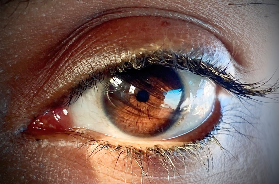
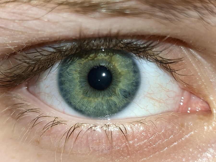
The posterior segment of the eye, encompassing the retina, choroid, and optic nerve, is a complex neural network integral to sight. Procedures performed in this region, while often life-changing for patients, also present unique considerations regarding the potential for prion transmission, albeit rare.
Surgical Interventions in the Posterior Segment
A range of surgical techniques are employed to address conditions affecting the posterior eye. These procedures, while varying in their invasiveness, share the commonality of entering the ocular environment.
Vitrectomy
Vitrectomy is a surgical procedure to remove the vitreous humor, the gel-like substance that fills the posterior cavity of the eye. It is commonly performed to treat conditions such as retinal detachment, diabetic retinopathy, macular hole, and floaters. During a vitrectomy, instruments are introduced through small incisions in the sclera to access and manipulate the vitreous and retinal structures. The surgical field involves direct contact with retinal tissues and the vitreous, which, in theory, could harbor prion proteins if a patient were unknowingly infected.
Retinal Detachment Repair
The surgical repair of retinal detachment involves reattaching the retina to the back wall of the eye. This can be accomplished through various techniques, including scleral buckling or vitrectomy, depending on the specific characteristics of the detachment. The goal is to prevent further damage to the photoreceptor cells and preserve or restore vision. As with vitrectomy, these procedures involve direct manipulation of retinal tissues.
Potential for Prion Contamination
The risk of prion transmission in the context of posterior eye procedures stems from the inherent persistence of prions and their presence in the central nervous system. While the eye is not considered central nervous system tissue itself, it is intricately connected to it via the optic nerve.
Access to Neural Tissue
Although direct access to the brain is not involved in standard posterior eye procedures, the optic nerve serves as a conduit for neural signals. The presence of PrPsc in the optic nerve, while not definitively established as a route of transmission for all prion diseases, represents a theoretical pathway for contamination. If PrPsc were present within the optic nerve head or surrounding tissues, instruments used in the posterior segment could potentially come into contact with it.
Instruments and Reusable Medical Devices
The primary concern for iatrogenic prion transmission via surgical instruments lies in their reusability. Standard sterilization methods, such as autoclaving, are not fully effective against prions. This means that if instruments are used on a patient with an undiagnosed prion disease, these instruments could retain infectious prion proteins. Subsequent use of these improperly decontaminated instruments on another patient could then lead to transmission. This is akin to a microscopic, yet incredibly durable, “sleeper agent” that lies dormant on surgical tools, waiting for its next opportunity to propagate.
Implantation of Devices
Procedures involving the implantation of intraocular lenses (IOLs) or other devices within the posterior segment also warrant consideration. While the primary risk associated with IOLs is bacterial or fungal infection, theoretical concerns arise if materials used in their manufacture were inadvertently contaminated during their production, though this is a highly unlikely scenario due to stringent manufacturing controls. The broader concern revolves around the instruments used for implantation.
Risk Assessment and Mitigation Strategies
The recognized risk of prion transmission through posterior eye procedures is exceptionally low, but the severity of prion diseases necessitates a rigorous approach to risk assessment and mitigation. This involves a multi-layered strategy encompassing patient screening, stringent decontamination protocols, and careful selection of surgical materials.
Pre-operative Screening and Patient History
While no definitive pre-operative test exists to screen all individuals for asymptomatic prion disease, a thorough patient history is a crucial first step. A detailed inquiry into neurological symptoms, family history of neurological disorders, and past medical procedures can help identify individuals at potentially higher risk.
Neurological Symptom Assessment
Any new-onset or progressing neurological symptoms, especially those affecting coordination, cognition, or vision, will raise a flag. These could be subtle at first, like a slight tremor or a growing forgetfulness, but they are critical indicators that demand further investigation. A careful physician will probe these symptoms deeply, much like a detective piecing together clues.
Family History and Genetic Predisposition
A known family history of prion diseases, such as familial CJD or FFI, immediately elevates a patient’s risk profile. This genetic predisposition means that the body’s own machinery for producing the prion protein may be subtly flawed, making it more susceptible to misfolding.
Past Medical Procedures
Inquiring about previous surgeries, especially those involving the nervous system or implantation of biological materials (like dura mater grafts), is important. The history of cadaver-derived growth hormone treatments in the past is also a critical piece of information.
Decontamination Protocols for Surgical Instruments
The persistence of prions against standard sterilization methods dictates the need for specialized decontamination protocols. These protocols are designed to be far more aggressive in their approach, aiming to break down or inactivate the infectious prion protein.
Chemical Disinfection
Several chemical agents have demonstrated efficacy in inactivating prions. Sodium hydroxide (NaOH) and sodium hypochlorite (bleach) are commonly used. These harsh chemicals work by denaturing the prion protein, altering its three-dimensional structure so it can no longer induce misfolding in normal proteins. The concentration and duration of exposure to these chemicals are critical factors in their effectiveness.
Thermal Inactivation
While autoclaving at standard temperatures may not entirely eliminate prions, elevated temperatures and extended exposure times can significantly reduce prion infectivity. Protocols often involve prolonged periods at higher temperatures (e.g., 134°C) under specific pressure conditions, creating a more formidable challenge for the resilient prion.
Enzymatic Cleaning
In some cases, enzymatic cleaning may be used in conjunction with other methods. Specific enzymes can help to break down proteins, and can be particularly effective in removing organic material that might shield prions from other decontamination agents.
The “Worst-Case Scenario” Approach
Medical institutions and regulatory bodies often adopt a “worst-case scenario” approach when it comes to prion decontamination in high-risk procedures. This means employing the most stringent protocols, even if the absolute risk is perceived as extremely low. It’s a philosophy of “better safe than sorry,” ensuring that every possible measure is taken.
Material Selection for Intraocular Devices
The materials used in intraocular devices and surgical instruments are subject to rigorous manufacturing and sterilization standards. The priority is to ensure that these materials themselves do not become a vector for prion transmission.
Single-Use Devices
The increasing use of single-use surgical instruments and disposable components for procedures like vitrectomy has significantly reduced the risk of iatrogenic prion transmission. Once used, these items are discarded, eliminating the need for reprocessing and the associated decontamination concerns. This is akin to discarding a potentially contaminated tool rather than attempting to purify and reuse it.
Material Sterilization Validation
For reusable devices, a critical aspect is the thorough validation of their sterilization processes. This involves demonstrating through rigorous testing that the chosen sterilization method effectively eliminates prion infectivity according to established guidelines. Every step of the manufacturing and sterilization process is meticulously documented and audited to ensure compliance.
The Extremely Low but Not Zero Risk
It is crucial to reiterate that the risk of acquiring a prion disease through posterior eye procedures is exceptionally low. The vast majority of individuals undergoing these procedures will never encounter such a complication. However, in medicine, even the smallest potential risk warrants careful consideration and robust preventative measures.
Rarity of Prion Diseases and Ocular Involvement
Prion diseases are rare conditions. Sporadic CJD affects approximately one to two people per million per year worldwide. Familial forms are even rarer, and iatrogenic transmission, while historically significant for certain procedures, is now extremely uncommon due to improved safety practices. Furthermore, documented evidence of prion replication or transmission specifically through the posterior segment of the eye in the absence of direct neurological involvement is scarce. The eye is not a primary site for prion accumulation in the same way the brain is.
Evolution of Safety Practices
The medical community has learned a great deal from past incidents of iatrogenic prion transmission. This learning has translated into evolution of safety practices, leading to a significant reduction in risk. The stringent decontamination protocols currently in place represent a direct response to this acquired knowledge.
The Importance of Transparency and Communication
Open and honest communication between healthcare providers and patients is paramount. Patients should feel empowered to ask questions about the potential risks and the measures in place to mitigate them. Understanding the rationale behind sterilization protocols and material choices can alleviate anxiety and foster trust. It’s about building a bridge of understanding, where both patient and doctor are on the same page when it comes to safeguarding health.
Recent discussions surrounding posterior eye procedures have raised concerns about the potential risk of prion transmission. A related article explores the implications of these procedures in detail, highlighting the need for stringent safety protocols to mitigate any possible risks. For further insights on this topic, you can read the article at Freaky Science, which delves into the intersection of medical practices and infectious disease transmission. Understanding these risks is crucial for both healthcare providers and patients alike.
What You Should Do
| Procedure | Prion Transmission Risk | Incidence Rate | Preventive Measures | Notes |
|---|---|---|---|---|
| Vitrectomy | Low to Moderate | Very rare; | Use of disposable instruments, thorough sterilization | Prion proteins may be present in vitreous body; risk theoretical but documented in rare cases |
| Retinal Detachment Repair | Low | Extremely rare | Strict sterilization protocols, single-use instruments when possible | Limited evidence of prion presence in retina; risk considered minimal |
| Posterior Capsulotomy | Very Low | No documented cases | Standard sterilization | Minimal tissue contact; prion transmission risk negligible |
| Intraocular Lens Implantation | Low | No documented cases | Use of sterile, single-use lenses and instruments | Prion contamination risk is theoretical |
| Endophthalmitis Treatment (Intravitreal Injection) | Very Low | No documented cases | Use of sterile, single-use syringes and needles | Prion transmission risk considered negligible |
As a patient, being informed is your most powerful tool. Taking an active role in your healthcare journey can lead to better outcomes and peace of mind.
Educate Yourself
This article serves as a starting point. Seek out reliable sources of information about posterior eye procedures and prion diseases. Do not rely on sensationalized or unverified accounts. Stick to established medical and scientific literature.
Discuss Concerns with Your Ophthalmologist
Before undergoing any posterior eye procedure, engage in a thorough discussion with your ophthalmologist. Do not hesitate to ask about:
- The specific procedure recommended for your condition.
- The rationale behind this recommendation.
- The potential risks and benefits.
- The hospital’s or clinic’s protocols for sterilization and infection control, particularly concerning prion diseases.
- Whether single-use instruments are utilized for your procedure.
- Any specific precautions they take due to the nature of your condition or the procedure itself.
Understand the Sterilization Procedures
While you are unlikely to be an expert in the intricacies of autoclave cycles or chemical concentrations, understanding that your healthcare provider adheres to stringent, validated protocols for instrument sterilization should be reassuring. Trust that the medical facility you are attending has invested in the necessary technology and training to ensure the highest level of safety.
Your Role in Patient History
Be forthright and comprehensive when providing your medical history. Any neurological concerns, family history of neurological conditions, or past medical treatments that might be relevant should be disclosed without reservation. Even seemingly minor details can be important pieces of the puzzle for your medical team.
By understanding the nature of prion diseases, the procedures involved, and the rigorous safety measures in place, you can approach posterior eye procedures with informed confidence. The medical community’s commitment to patient safety is an ongoing endeavor, constantly adapting and improving based on scientific understanding and past experiences. Your active participation in this process is a vital component of ensuring your well-being.
FAQs
What are posterior eye procedures?
Posterior eye procedures refer to surgical or medical interventions performed on the back part of the eye, including the retina, vitreous, and optic nerve. Common examples include vitrectomy, retinal detachment repair, and treatment for macular degeneration.
What is prion disease and how is it related to eye procedures?
Prion diseases are a group of rare, fatal neurodegenerative disorders caused by misfolded prion proteins. They can potentially be transmitted through contaminated surgical instruments. In the context of eye procedures, there is concern about the risk of transmitting prion diseases such as Creutzfeldt-Jakob disease (CJD) if instruments are not properly sterilized.
Is there a known risk of prion transmission during posterior eye surgeries?
While prion transmission through posterior eye surgeries is extremely rare, it is theoretically possible if contaminated instruments are reused without adequate sterilization. The vitreous and retina are considered tissues with low to moderate infectivity, so strict sterilization protocols are essential to minimize any risk.
What sterilization measures are recommended to reduce prion risk in eye surgeries?
To reduce prion transmission risk, surgical instruments used in posterior eye procedures should undergo rigorous sterilization processes, including extended autoclaving at high temperatures and the use of chemical disinfectants effective against prions. Some guidelines recommend using disposable instruments or dedicating specific sets for high-risk procedures.
Should patients be concerned about prion risk when undergoing posterior eye procedures?
The risk of prion transmission during posterior eye procedures is extremely low due to stringent sterilization standards and infection control protocols. Patients should discuss any concerns with their ophthalmologist, but in general, posterior eye surgeries are considered safe with respect to prion disease transmission.