You are standing on the precipice of discovery, a cosmic cartographer charting unseen landscapes. The soil beneath your feet, seemingly inert, is a bustling metropolis, teeming with life invisible to the naked eye. Among these microscopic denizens, a select group, the mirror microbes, holds a particular allure. They are nature’s serendipitous reflections, organisms whose metabolic processes echo the characteristics of specific mineral compounds. Unlocking their secrets can illuminate pathways for bioremediation, biomining, and a deeper understanding of biogeochemical cycles. This guide is your compass, designed to equip you with the knowledge and techniques to venture into this hidden realm and detect these fascinating mirror microbes.
Mirror microbes are a fascinating niche within the vast microbial kingdom. Their defining characteristic is their ability to mimic or interact with specific mineral phases in a way that is diagnostically useful. This isn’t a conscious act of imitation, but rather a consequence of their evolutionary adaptation to thrive in environments rich in certain elements or compounds. Think of them as tiny, biological alchemists, their enzymes and metabolic byproducts acting as keys that unlock or subtly alter the rocks and minerals around them.
The Biological Mimicry Explained
At the heart of mirror microbe identification lies the concept of biological mimicry. These microorganisms have evolved to produce extracellular enzymes or metabolites that can dissolve, precipitate, or otherwise alter mineral surfaces. For example, a microbe might excrete an organic acid that chelters metal ions from a sulfide mineral, effectively “dissolving” it in a biological sense, mimicking the natural weathering processes of that mineral. Conversely, another microbe might create conditions that favor the precipitation of a particular mineral phase from dissolved ions in the soil pore water. This interaction is crucial because it can influence mineral formation, erosion, and the availability of essential nutrients.
Why Detect Mirror Microbes? Applications in Science and Industry
The detection of mirror microbes is far more than an academic exercise. Their unique abilities offer tangible solutions to pressing global challenges and open new avenues for scientific exploration.
Bioremediation: Nature’s Cleanup Crew
One of the most significant applications lies in bioremediation. Certain mirror microbes can be harnessed to clean up contaminated sites. For instance, microbes that metabolize or sequester heavy metals can be used to extract these toxic elements from polluted soils or wastewater. They are the silent janitors of the ecosystem, tirelessly working to restore balance. This is particularly relevant in areas affected by industrial mining or spills, where heavy metal contamination poses a severe threat to environmental and human health.
Biomining: Extracting Value from Earth
Biomining, also known as bioleaching, is another area where mirror microbes shine. These organisms can be employed to extract valuable metals from low-grade ores. Instead of energy-intensive physical and chemical processes, low concentrations of certain metals, such as copper or gold, can be solubilized by microbial action. This provides a more environmentally friendly and economically viable approach to resource extraction. Imagine these microbes as tiny, efficient prospectors, sifting through the earth for hidden treasures.
Understanding Geochemical Cycles
Beyond practical applications, detecting mirror microbes provides invaluable insights into fundamental biogeochemical cycles. Their interactions with minerals play a significant role in nutrient cycling, rock weathering, and the formation of sedimentary rocks over geological timescales. By understanding these microbial drivers, you gain a deeper appreciation for the dynamic processes that shape our planet. They are the microscopic gears in the colossal engine of Earth’s geology.
Detecting mirror microbes in soil samples is a fascinating area of research that can provide insights into soil health and ecosystem dynamics. For those interested in learning more about innovative techniques and methodologies in microbial detection, a related article can be found at Freaky Science. This resource offers valuable information on the latest advancements in soil microbiology, including the use of molecular techniques and bioinformatics to identify and analyze microbial communities.
Sampling Strategies: Gathering Your Microbial Treasures
The journey to detecting mirror microbes begins with careful and strategic sampling. The soil is a heterogeneous environment, and your approach will dictate the success of your endeavor. Think of soil sampling as carefully unearthing ancient artifacts; you need the right tools and techniques to bring hidden histories to light.
Defining Your Target Mineral(s)
Before you even set foot in the field, you must clearly define which mineral or suite of minerals you are interested in. Are you looking for microbes associated with iron sulfides, for example, or perhaps those that interact with carbonate minerals? Your choice of target mineral will guide your sampling locations and the subsequent analytical methods. This initial focus is like honing your telescope on a specific celestial body before embarking on an astronomical survey.
Representative Soil Collection
Obtaining representative soil samples is paramount. Avoid sampling from disturbed areas, such as the edges of trails or freshly dug pits, as these may not reflect the natural microbial communities. Aim for areas that are undisturbed and reflect the geological and environmental conditions associated with your target mineral.
Depth and Stratification
Consider the depth at which you are sampling. Microbial communities can vary significantly with soil depth due to differences in oxygen availability, moisture content, and the proximity to mineral sources. If your target mineral is typically found at a specific depth, focus your sampling efforts there. You might also consider collecting samples from multiple depths to capture a broader picture of microbial stratification.
Sample Size and Replication
The size of your samples should be sufficient to capture the microbial diversity present. Standard soil sampling protocols often recommend collecting samples from several points within a defined area and then pooling them to create a composite sample. Replication is also essential; taking multiple independent samples from the same location will allow you to assess the variability and robustness of your findings.
Field Preservation of Samples
Once collected, your precious microbial samples need proper preservation to prevent unwanted microbial activity and degradation. This is akin to carefully preserving delicate specimens for later study.
Temperature Control
Immediately after collection, samples should be kept cool. Refrigeration (4°C) is generally recommended for short-term storage, as it slows down microbial metabolism without permanently damaging cells. For longer-term storage, freezing (-20°C or -80°C) is often necessary. Ensure your freezing and thawing protocols are gentle to minimize cell lysis.
Preventing Contamination
Sterility is your ally in this endeavor. Use clean sampling tools (sterilized beforehand) and containers to prevent the introduction of foreign microbes. Work in a clean environment, ideally under a laminar flow hood if possible, to minimize airborne contamination.
Isolation and Cultivation: Orchestrating Microbial Growth
With your carefully collected samples in hand, the next step is to coax the mirror microbes out of their native environment and into a controlled setting where you can study them. This is where you become a microbial gardener, carefully tending to the seeds of life.
Culturability: The Art of the Possible
It is important to acknowledge that not all microbes are easily culturable in a laboratory setting. Many possess highly specific nutritional requirements or exist in symbiotic relationships that are difficult to replicate. Therefore, your success in isolating mirror microbes will depend on your ability to design appropriate growth media.
Designing Selective Media
The key to isolating specific microbes lies in designing selective growth media. These media are formulated to provide nutrients that favor the growth of your target microbes while inhibiting the growth of others. This is your net, cast into the microbial sea to catch your specific quarry.
Nutrient Selection
Consider the likely metabolic pathways and nutritional needs of your target mirror microbes. If you are looking for iron-oxidizing bacteria, your media might be low in readily available organic carbon but rich in iron sources. Conversely, if you are targeting sulfate-reducing bacteria, sulfur compounds would be essential components.
pH and Other Environmental Factors
Adjusting the pH of the media is crucial, as different microbes thrive at different pH ranges. Similarly, consider the need for specific electron donors or acceptors relevant to your target microbes’ metabolism. For example, in some cases, you might need to create anaerobic conditions to cultivate strictly anaerobic microorganisms.
Incubation Conditions: Simulating the Soil Environment
Once your selective media is prepared and inoculated, you must provide the right incubation conditions to encourage microbial growth. This is your controlled greenhouse.
Temperature and Atmosphere
Maintain incubation temperatures that are physiologically suitable for the microbes you expect to find. This is often close to the ambient temperature of the soil from which the sample was taken. Similarly, consider the atmospheric requirements – aerobic, microaerobic, or anaerobic conditions may be necessary.
Time for Growth
Microbial growth can be slow, especially for specialized organisms. Be patient and allow sufficient time for colonies to develop. Regular monitoring of your cultures is essential to detect the first signs of growth.
Detection and Characterization: Unmasking the Mirror
Once you have obtained microbial growth, the challenge shifts to identifying and characterizing the specific mirror microbes within your cultures. This is where you put on your detective hat and begin to unmask the hidden identities.
Phenotypic and Macroscopic Observations
Your initial observations can provide valuable clues. Look for characteristic colony morphologies, growth patterns, and any observable changes in the surrounding media.
Pigmentation and Colony Morphology
Some microbial colonies exhibit distinct colors due to pigment production, which can be indicative of certain metabolic activities. The shape, size, and texture of colonies can also be identifying features.
Changes in the Surrounding Media
Observe any color changes, precipitate formation, or gas production in the media around the growing colonies. These secondary effects can be direct evidence of the microbes’ interaction with the media components, which may mirror their interactions with minerals in the soil.
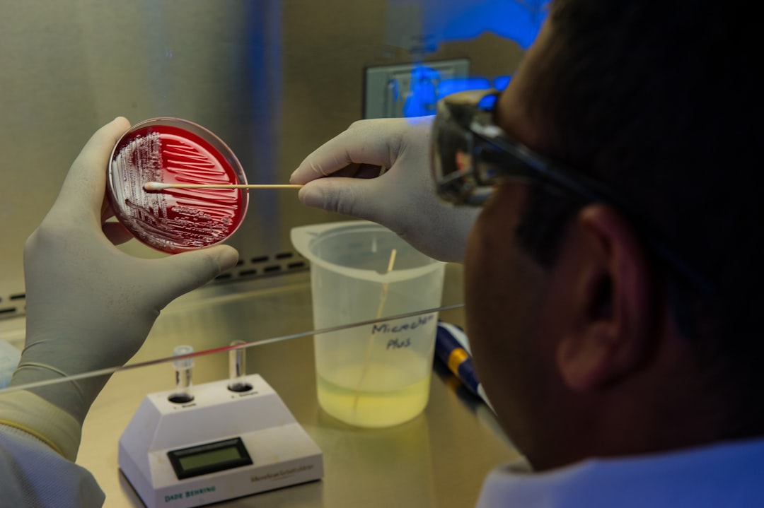
Microscopy: A Glimpse into the Microbial World
Microscopy is an indispensable tool for visualizing and characterizing individual microbial cells.
Gram Staining and Other Differential Stains
Gram staining is a fundamental technique that classifies bacteria based on their cell wall composition. Other differential stains can highlight specific cellular structures or inclusions.
Phase Contrast and Darkfield Microscopy
These techniques improve the visibility of unstained, living microorganisms, allowing you to observe their motility and general morphology without the need for harsh staining procedures.
Electron Microscopy (TEM and SEM)
For detailed ultrastructural analysis, Transmission Electron Microscopy (TEM) and Scanning Electron Microscopy (SEM) are invaluable. SEM can reveal the surface topography of the microbes and their interactions with mineral particles that might have been present on your isolation media. TEM provides cross-sectional views of cellular structures.
Biochemical and Molecular Techniques: The Ultimate Identification
While phenotypic and microscopic observations are important, biochemical and molecular techniques offer the most definitive identification of mirror microbes. These methods delve into the molecular blueprint of the organism.
Biochemical Assays
A range of biochemical tests can be performed to determine the metabolic capabilities of your isolates. This includes testing for the presence of specific enzymes, carbon source utilization, and the production of various metabolic end-products.
DNA Sequencing: The Microbial Fingerprint
The gold standard for microbial identification is DNA sequencing. By sequencing specific genes, such as the 16S ribosomal RNA (rRNA) gene, you can generate a unique genetic fingerprint for each isolate. This sequence can then be compared to databases of known microbial genomes to identify the organism’s taxonomic affiliation.
Polymerase Chain Reaction (PCR)
PCR is a technique used to amplify specific DNA sequences, allowing you to obtain enough DNA for sequencing from even small samples. You will likely employ PCR to amplify the 16S rRNA gene from your isolated microbes.
Sanger Sequencing and Next-Generation Sequencing (NGS)
Sanger sequencing is a well-established method for sequencing individual DNA fragments. For a broader understanding of microbial community composition and to explore the functional potential of your isolates, Next-Generation Sequencing (NGS) platforms offer high-throughput sequencing capabilities.
Mineral Interaction Assays: Confirming the “Mirror”
The ultimate confirmation of a mirror microbe lies in demonstrating its direct interaction with your target mineral. This step is the crucial handshake between the microbe and its mineral counterpart.
Batch Culture Assays
In batch culture assays, you would expose purified isolates to sterile mineral powders or solutions containing relevant ions. You then monitor for changes in the mineral phase (e.g., dissolution, precipitation) or changes in the microbial population over time. Techniques like X-ray Diffraction (XRD) can be used to identify mineral phases formed or altered, while Inductively Coupled Plasma Mass Spectrometry (ICP-MS) can quantify dissolved metals.
Biofilm Formation and Mineral Colonization Studies
Observe if your isolates readily form biofilms on mineral surfaces. This can be assessed using microscopy and staining techniques. The ability of microbes to adhere to and colonize specific minerals is a strong indicator of a potential mirror microbe.
Detecting mirror microbes in soil samples is a fascinating area of research that can provide insights into soil health and ecosystem dynamics. For those interested in exploring this topic further, a related article discusses innovative techniques and methodologies for microbial detection, which can enhance our understanding of soil microbiomes. You can read more about these techniques in this informative piece on Freaky Science. This resource offers valuable information that complements the study of mirror microbes and their implications for agriculture and environmental science.
Troubleshooting and Advanced Techniques: Refining Your Search
| Method | Description | Key Metrics | Advantages | Limitations |
|---|---|---|---|---|
| Microscopic Examination | Direct observation of soil microbes using light or electron microscopy | Cell morphology, size, and abundance | Quick visualization, identifies cell structure | Cannot differentiate mirror microbes specifically, low resolution for species ID |
| Fluorescence In Situ Hybridization (FISH) | Use of fluorescent probes targeting specific microbial RNA sequences | Fluorescence intensity, probe binding specificity | Specific detection of target microbes, spatial distribution | Requires known target sequences, limited by probe design |
| Polymerase Chain Reaction (PCR) | Amplification of DNA sequences specific to mirror microbes | Cycle threshold (Ct) values, amplicon size | Highly sensitive and specific, detects low abundance microbes | Requires prior knowledge of target DNA, risk of contamination |
| Metagenomic Sequencing | Sequencing all microbial DNA in soil to identify mirror microbes | Read counts, sequence coverage, diversity indices | Comprehensive profiling, detects unknown microbes | Expensive, complex data analysis |
| Culture-Based Methods | Growing microbes on selective media to isolate mirror microbes | Colony forming units (CFU), growth rate | Allows functional studies, isolates live microbes | Many microbes are unculturable, time-consuming |
| Stable Isotope Probing (SIP) | Tracking incorporation of labeled substrates into microbial DNA/RNA | Isotope enrichment levels, labeled nucleic acid recovery | Links function to identity, detects active microbes | Requires specialized equipment, costly |
Your journey may not always be smooth. Expect challenges and be prepared to adapt your strategies. The path of discovery is often paved with a few bumps.
Dealing with Unsuccessful Cultivation
If you are repeatedly failing to cultivate your target microbes, consider the possibility that your isolation media and conditions may not be optimal.
Exploring Novel Media Formulations
Experiment with a wider range of nutrient sources, inorganic salts, and buffering agents. Consider using environmental samples directly as inocula into your enriched media.
Utilizing Environmental Mimicry
Try to recreate the specific soil conditions, such as pore water chemistry and gas composition, as closely as possible in your laboratory media.
Advanced Spectroscopic and Imaging Techniques
For more sophisticated characterization of microbial-mineral interactions, advanced techniques can provide deeper insights.
Atomic Force Microscopy (AFM)
AFM can provide high-resolution topographic imaging of mineral surfaces and microbial cells, allowing you to visualize direct interactions at the nanoscale without the need for extensive sample preparation.
Raman Spectroscopy
Raman spectroscopy can identify the molecular composition of both microbes and minerals, providing a non-destructive way to study their interactions and transformations in situ.
Metagenomics and Metatranscriptomics
For studying unculturable microbes or entire microbial communities, metagenomics (studying all the DNA in a sample) and metatranscriptomics (studying all the RNA) are powerful tools. These techniques allow you to infer the presence and functional potential of mirror microbes directly from environmental samples, bypassing the need for cultivation.
Isotopes and Tracers
The use of stable isotopes or radioactive tracers can reveal metabolic pathways and element cycling associated with mirror microbes. By labeling specific elements in your culture media or soil samples, you can track their movement and transformation by the microbes.
Your quest to detect mirror microbes is a testament to your curiosity and your willingness to peer into the unseen. Each step, from careful sampling to sophisticated molecular analysis, brings you closer to understanding these remarkable organisms. The soil, a universe in miniature, holds its secrets close, but with the right tools and a persistent spirit, you can indeed become a successful cartographer of its microbial inhabitants, revealing the hidden reflections that shape our world.
FAQs
What are mirror microbes in soil samples?
Mirror microbes refer to microorganisms that exhibit symmetrical or reflective characteristics in their structure or behavior, often identified through specific staining or imaging techniques in soil microbiology.
Why is it important to detect mirror microbes in soil samples?
Detecting mirror microbes helps in understanding soil health, microbial diversity, and ecological functions, which can impact agriculture, environmental monitoring, and bioremediation efforts.
What methods are commonly used to detect mirror microbes in soil?
Common methods include microscopy techniques such as fluorescence microscopy, scanning electron microscopy (SEM), and molecular approaches like DNA sequencing and specific staining protocols to highlight mirror-like features.
How should soil samples be prepared for detecting mirror microbes?
Soil samples should be collected aseptically, homogenized, and may require dilution or suspension in a buffer solution. Proper fixation and staining are often necessary before microscopic examination.
Can mirror microbes be cultured in the laboratory for further study?
Some mirror microbes can be cultured using selective media and controlled environmental conditions, but many soil microbes are unculturable and require molecular or imaging techniques for detection and analysis.