Perception, for you, is not a passive reception of sensory data; it is an active construction, a complex interplay between your sensory organs and your brain. You don’t just “see” the world; you interpret it, filter it, and imbue it with meaning based on your past experiences, expectations, and even your current emotional state. This intricate process, often taken for granted in your daily life, is the subject of intense investigation within neuroscience. This article will delve into the mechanisms by which you, as a human being, perceive the world around you, focusing specifically on visual perception, a primary gateway to your understanding of your environment.
To understand how you perceive, you must first understand the initial conduit of visual information: your eye. Think of your eye not as a simple window, but as a sophisticated, self-adjusting biological camera. You can learn more about split brain consciousness in this informative video.
The Retina: The Brain’s Outer Layer
At the back of your eye, you find the retina, a delicate, multi-layered sheet of neural tissue. This isn’t just a screen; it’s an extension of your brain, containing the photoreceptor cells responsible for converting light into electrical signals.
Rods and Cones: Your Photodetectors
Within the retina, you possess two primary types of photoreceptor cells: rods and cones.
- Rods: These highly sensitive cells are your primary detectors for dim light and peripheral vision. They are crucial for your night vision, allowing you to discern shapes and movement in low-light conditions. However, rods are monochromatic, meaning they do not contribute to your perception of color. You have a significantly larger number of rods than cones, underscoring their importance in basic visual detection.
- Cones: These cells are responsible for your high-acuity vision and your perception of color. They require brighter light to function optimally. You possess three types of cones, each sensitive to different wavelengths of light: short (blue), medium (green), and long (red). The combination of signals from these three cone types allows you to perceive the vast spectrum of colors you experience. The fovea, a small central pit in your retina, is densely packed with cones, providing you with your sharpest, most detailed vision.
Retinal Ganglion Cells: The Information Converters
Before electrical signals leave your eye, they are processed by a network of interneurons within the retina, ultimately converging on retinal ganglion cells. Each retinal ganglion cell receives input from multiple photoreceptors, consolidating and encoding visual information into action potentials. These cells are specialized to detect specific features, such as contrasts, edges, and movement. Their axons then bundle together to form the optic nerve.
Understanding how perception works in neuroscience is a fascinating topic that delves into the intricate processes by which our brains interpret sensory information. For a deeper exploration of this subject, you can read a related article that discusses the mechanisms of perception and the role of neural pathways in shaping our experiences. This article provides valuable insights into how our brains construct reality based on the information received from our senses. To learn more, visit this article.
The Optic Nerve and Beyond: The Visual Pathway
Once the visual information has been transduced and initially processed by your retina, it embarks on a complex journey through your brain along the visual pathway.
The Optic Chiasm: A Crossroads of Information
The optic nerve from each of your eyes travels to the optic chiasm, a crucial point where fibers partially cross over. This decussation ensures that visual information from your left visual field (seen by both eyes) is processed by your right cerebral hemisphere, and information from your right visual field is processed by your left hemisphere. This anatomical arrangement is fundamental to your unified perception of the world.
The Lateral Geniculate Nucleus (LGN): The Thalamic Relay Station
From the optic chiasm, the majority of visual fibers project to the lateral geniculate nucleus (LGN) in your thalamus. The LGN acts as a sophisticated relay station, not merely passing information along but also performing further processing and modulation. It receives feedback from other brain regions, indicating that ascending visual information is not a one-way street, but subject to top-down influence even at this early stage. The LGN is organized into distinct layers, each processing specific types of visual information, such as color, motion, or form.
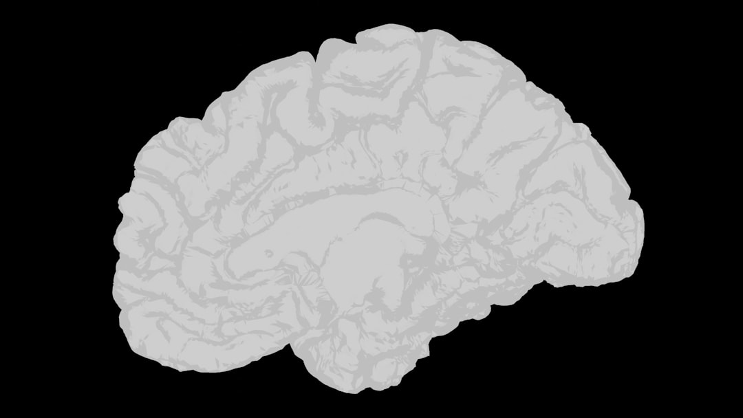
The Primary Visual Cortex (V1): The Genesis of Conscious Sight
The LGN projects to the primary visual cortex, often referred to as V1 or the striate cortex, located in the occipital lobe at the back of your brain. This region is the initial destination for consciously perceived visual information.
Orientation Selectivity: Building Blocks of Form
In V1, you encounter neurons that are exquisitely sensitive to specific orientations of lines or edges. These “simple cells” and “complex cells” fire vigorously when presented with stimuli like a vertical line, but remain silent for a horizontal one. This specialization is one of the earliest steps in your brain’s construction of form and shape. Think of it as your brain assembling individual brushstrokes into a coherent image.
Ocular Dominance Columns: Integrating Binocular Input
V1 is also organized into ocular dominance columns, with neurons in alternating columns preferentially responding to input from one eye or the other. This columnar organization is crucial for integrating the slightly different images received by your two eyes, providing depth perception.
Beyond V1: The Dorsal and Ventral Streams

After initial processing in V1, visual information diverges into two major processing streams, often referred to as the “what” and “where” pathways, or the ventral and dorsal streams, respectively. This specialization allows your brain to simultaneously process different aspects of the visual scene.
The Ventral Stream: Unraveling “What” You See
The ventral stream, originating in V1 and extending into your temporal lobe, is primarily responsible for object recognition – for identifying what you are looking at.
Fusiform Face Area: The Face Detector
Within your temporal lobe, a region known as the fusiform face area (FFA) exhibits remarkable selectivity for faces. Damage to this area can lead to prosopagnosia, a condition where individuals struggle to recognize familiar faces, highlighting the specialized nature of this cortical region. This suggests that your brain has evolved dedicated mechanisms for identifying this crucial social stimulus.
Parahippocampal Place Area: Recognizing Your Surroundings
Adjacent to the FFA, the parahippocampal place area (PPA) shows preferential activation to scenes and places. This area is critical for your ability to orient yourself within an environment and recognize familiar locations. When you enter a room and immediately categorize it as a “kitchen” or a “forest,” your PPA is actively involved.
Object Recognition: A Hierarchical Process
Your brain doesn’t recognize objects all at once. The ventral stream employs a hierarchical processing model. Simpler features like edges and corners are detected in earlier visual areas, and these are then combined into more complex shapes, ultimately leading to the recognition of entire objects. This process is remarkably robust, allowing you to recognize an object regardless of its size, position, or lighting conditions. Think of it as a sculptor building a statue: starting with rough blocks and progressively refining details.
The Dorsal Stream: Navigating “Where” and “How”
The dorsal stream, originating in V1 and extending into your parietal lobe, is involved in processing spatial information, motion, and guiding your actions. It answers the questions of where an object is and how you can interact with it.
Motion Perception: Tracking Moving Objects
Specialized areas within the dorsal stream are dedicated to perceiving motion. An area known as V5 or MT (middle temporal area) is particularly crucial for detecting and interpreting the movement of objects in your visual field. Damage to this area can lead to akinetopsia, a condition where individuals perceive the world as a series of still frames rather than continuous motion.
Spatial Navigation: Your Internal GPS
The dorsal stream plays a vital role in your ability to navigate your environment. It constantly processes information about your own body’s position relative to objects around you, enabling you to reach for a cup or walk around an obstacle without conscious effort. This area effectively acts as your brain’s internal GPS, constantly updating your location and the location of objects around you.
Visuomotor Control: Guiding Your Actions
This stream is intimately connected with your motor systems, allowing you to accurately reach for, grasp, or manipulate objects. When you throw a ball, catch a frisbee, or simply pick up a pen, the dorsal stream is actively ensuring your movements are precisely coordinated with the visual information you are receiving.
Top-Down Processing: The Brain’s Predictive Power

While sensory signals travel from your eyes to your brain (bottom-up processing), your brain also actively influences what you perceive through top-down processing. This means your prior knowledge, expectations, and goals profoundly shape your visual experience.
Expectation and Attention: Shaping Your Reality
If you expect to see something, you are more likely to perceive it, even with ambiguous sensory input. Attention, too, acts as a spotlight, enhancing the processing of relevant stimuli and suppressing irrelevant ones. When you are searching for your keys, your visual system is biased towards detecting key-like shapes, making you more likely to spot them amidst clutter. This illustrates that what you perceive is not merely what is “out there,” but also what your brain is “looking for.”
Perceptual Constancies: A Stable World
Despite constant changes in the sensory input your eyes receive (e.g., an object moving further away, changing illumination), you perceive a stable and consistent world. This is due to perceptual constancies.
Size Constancy: Objects Don’t Shrink
Even as an object moves closer or further away, you perceive its size as constant. Your brain compensates for the changing retinal image size by factoring in depth cues. You instinctively understand that a car approaching you is not growing larger, but simply getting closer.
Shape Constancy: Recognizing Objects from Various Angles
You recognize objects even when viewed from different angles, which alters their retinal image. A door remains a door whether it’s closed, partially open, or seen from an oblique angle. Your brain is adept at reconstructing the true, invariant shape of an object despite these variations.
Brightness and Color Constancy: Consistent Illumination
The perceived brightness and color of an object remain relatively consistent despite changes in the ambient lighting. A red apple appears red under bright sunlight, fluorescent light, or in dim conditions, even though the wavelengths of light reflecting off it are significantly different. Your brain adjusts for the perceived illuminant to maintain a stable color experience.
Understanding how perception works in neuroscience is a fascinating journey into the complexities of the human brain. Recent studies have shown that our sensory experiences are not merely reflections of the external world but are significantly shaped by our brain’s interpretations and prior knowledge. For those interested in exploring this topic further, an insightful article on the subject can be found at Freaky Science, which delves into the intricate mechanisms behind perception and how they influence our interactions with the world around us.
The Illusion of Sight: When Perception Deviates from Reality
| Metric | Description | Typical Value/Range | Relevance to Perception |
|---|---|---|---|
| Neural Firing Rate | Frequency at which neurons fire action potentials | 0-200 Hz | Encodes sensory information intensity and timing |
| Latency of Sensory Response | Time delay between stimulus onset and neural response | 10-100 ms depending on modality | Determines speed of perception processing |
| Receptive Field Size | Area of sensory space that activates a neuron | Micrometers to centimeters (varies by sensory system) | Influences spatial resolution of perception |
| Synaptic Plasticity Rate | Rate of change in synaptic strength | Milliseconds to hours | Supports learning and adaptation in perception |
| Oscillation Frequency | Brain wave frequency bands involved in perception | Delta (1-4 Hz), Theta (4-8 Hz), Alpha (8-12 Hz), Beta (12-30 Hz), Gamma (30-100 Hz) | Coordinates neural activity during sensory processing |
| Signal-to-Noise Ratio (SNR) | Ratio of meaningful sensory signal to background noise | Varies widely; higher SNR improves perception accuracy | Critical for distinguishing stimuli in noisy environments |
| Perceptual Threshold | Minimum stimulus intensity detectable by the sensory system | Varies by modality; e.g., visual threshold ~1 photon | Defines sensitivity limits of perception |
The constructive nature of perception is most evident in visual illusions. These phenomena demonstrate how your brain’s inference-making processes can sometimes lead to perceptions that don’t precisely match the physical reality.
Optimal Guesswork: The Brain’s Best Bet
Many illusions arise because your brain is constantly making “optimal guesses” about the world based on ambiguous sensory input. It uses heuristics and shortcuts to rapidly interpret scenes, and sometimes these shortcuts lead to misinterpretations. For example, the Müller-Lyer illusion, where two lines of equal length appear different due to the direction of their arrowheads, is thought to stem from your brain’s application of depth cues to flat images.
The Blind Spot: Evidence of Filling In
You possess a “blind spot” in each eye, a region where the optic nerve exits the retina and there are no photoreceptors. Yet, you are almost never aware of it. Your brain “fills in” the missing information using surrounding visual cues and expectations, creating a seamless perceptual experience. This is a powerful illustration of your brain actively constructing your reality rather than passively receiving it.
In conclusion, your ability to see is not a simple act of registering light, but a sophisticated, multi-stage process of transduction, transmission, and extensive neural computation. From the initial capture of light by your photoreceptors to the intricate dance of neurons in your visual cortex, and the powerful influence of your expectations and prior knowledge, your brain actively constructs the rich, detailed, and coherent visual world that you experience every moment of your waking life. Understanding these processes provides a profound insight into the very nature of your reality.
FAQs
What is perception in neuroscience?
Perception in neuroscience refers to the process by which the brain interprets sensory information from the environment to form an understanding or awareness of objects, events, and spatial relationships.
How does the brain process sensory information?
The brain processes sensory information by receiving signals from sensory organs, such as the eyes, ears, and skin, which are then transmitted to specific areas of the brain for interpretation and integration into a coherent perceptual experience.
What role do neurons play in perception?
Neurons transmit electrical and chemical signals that carry sensory information to the brain. They also participate in complex networks that analyze and interpret these signals, enabling perception to occur.
Can perception be influenced by prior knowledge or expectations?
Yes, perception is influenced by prior knowledge, experiences, and expectations, which can shape how sensory information is interpreted, leading to phenomena such as perceptual illusions or biases.
What are some common methods used to study perception in neuroscience?
Common methods include brain imaging techniques like fMRI and EEG, electrophysiological recordings, behavioral experiments, and computational modeling to understand how sensory information is processed and perceived.