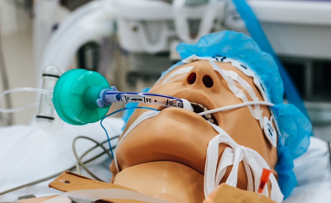
You’ve just undergone surgery. The last thing you remember is the gentle hum of the operating room, the masked faces, and then… nothing. You wake up feeling groggy, disoriented, and with a faint memory of a dreamless void. But what was actually happening inside your brain during that period of unconsciousness? This is where electroencephalography, or EEG, steps in, offering a fascinating glimpse into the hidden world of brain activity under anesthesia.
Your brain is a constant hive of electrical activity, a complex network of neurons firing and communicating. This electrochemical chatter generates tiny electrical signals that can be detected on the scalp. EEG is the technology that picks up these signals, translating them into wavy lines on a screen, each representing a different pattern of brain activity. These patterns, or brain waves, are categorized by their frequency and amplitude, and they tell a story about your mental state.
The Spectrum of Consciousness: From Awake to Asleep
You’re probably familiar with the idea of brain waves like alpha and theta. When you’re awake and relaxed, your brain often hums with alpha waves, a rhythmic beat associated with a state of calm alertness. As you drift off to sleep, these alpha waves slow down and are replaced by theta waves, indicative of deeper relaxation. This spectrum of brain wave activity is well-understood and forms the baseline against which we can compare what happens during anesthesia.
The EEG Machine: Your Window to the Mind
The EEG machine itself is a marvel of non-invasive technology. Small, metal discs called electrodes are attached to your scalp using a conductive paste. These electrodes are connected by wires to an amplifier and recording device, which then processes and displays the electrical signals. For you, the patient, this process is completely painless and requires no preparation beyond what’s needed for your surgery. It’s a passive observation, allowing scientists and clinicians to monitor the intricate workings of your brain without any interference.
Electroencephalography (EEG) provides valuable insights into brain activity during anesthesia, revealing how different anesthetic agents affect neural oscillations and connectivity. For a deeper understanding of these effects, you can explore a related article that discusses the implications of EEG monitoring in anesthesia practices. This article delves into the relationship between EEG patterns and the depth of anesthesia, highlighting the importance of real-time monitoring for patient safety. To read more, visit this article.
Anesthesia: A Controlled Descent into Unconsciousness
Anesthesia is a complex medical intervention designed to block pain and induce a state of unconsciousness, allowing surgical procedures to be performed safely and comfortably. It’s not simply turning off the brain; rather, it’s a carefully orchestrated manipulation of brain activity, and EEG is the primary tool for observing and understanding this manipulation.
The Art and Science of Anesthesia Administration
Administering anesthesia is both an art and a science. Anesthesiologists use a combination of drugs – inhaled agents, intravenous medications – to achieve the desired level of unconsciousness and analgesia. The precise dosage and combination are tailored to the individual patient, the type of surgery, and the duration of the procedure. Historically, anesthesiologists relied on subtle cues like breathing patterns, heart rate, and muscle tone to gauge a patient’s depth of anesthesia. However, EEG provides an objective, quantifiable measure.
The Goal: Reversible Unconsciousness
The key characteristic of a safe anesthetic is its reversibility. The goal is to induce a state where you are unaware and pain-free, but can be reliably brought back to consciousness once the procedure is complete. Understanding how anesthetic agents affect brain wave patterns is crucial for ensuring this reversibility and for monitoring the safety and efficacy of the anesthetic.
EEG Signatures of Anesthesia: A Shifting Landscape
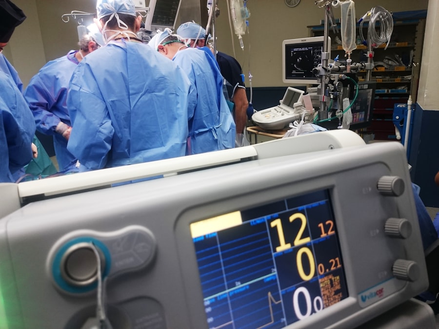
When you are given anesthetic agents, your brain’s electrical activity undergoes profound changes. EEG reveals these alterations as distinct shifts in the characteristic brain wave patterns. It’s like observing a symphony orchestra that gradually transforms its music, moving from lively allegro to a slow, deliberate adagio.
The Dominance of Slow Waves: A hallmark of Deep Sleep
One of the most prominent changes you’ll observe on an EEG trace during anesthesia is the emergence of slow, high-amplitude waves. These are often delta waves (typically below 4 Hz) and slow theta waves. The appearance of these slow waves is a strong indicator that your brain is entering a state of deep unconsciousness. It’s thought that these slow oscillations reflect a widespread disruption of communication between different brain regions, a necessary component for suppressing consciousness and perception.
Burst Suppression: Periods of Silence
In deeper levels of anesthesia, you might even see a phenomenon called “burst suppression.” This is characterized by periods of electrical silence – the brain waves virtually flatlining – interspersed with brief bursts of electrical activity. This pattern suggests that large areas of the brain are temporarily silenced, only to regain some sporadic activity before falling silent again. While it might sound alarming, burst suppression can be a sign of effective deep anesthesia, ensuring that no pain signals are being processed. However, anesthesiologists carefully monitor this to avoid prolonged periods of silence, which could indicate excessive anesthetic depth.
The Impact of Different Anesthetic Agents
The specific EEG patterns observed can also vary depending on the type of anesthetic agent used. For example, inhaled anesthetics like sevoflurane and isoflurane tend to induce a more prominent slow-wave activity and can lead to burst suppression at higher concentrations. Intravenous agents, such as propofol, can also induce slow waves but might have different characteristics in their effects on frontal lobe activity, for instance. Understanding these nuances allows anesthesiologists to fine-tune their anesthetic strategy based on the EEG feedback.
Beyond Unconsciousness: Monitoring Brain State and Awareness
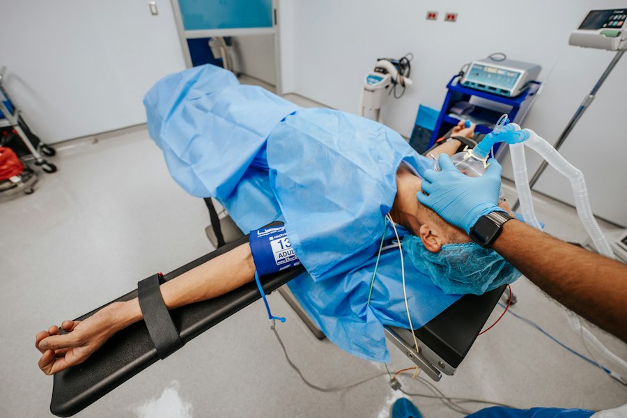
While the primary goal of anesthesia is unconsciousness, EEG provides more than just a binary on-off switch. It can help clinicians monitor the depth of anesthesia and, crucially, detect potential signs of awareness under anesthesia.
The Depth of Sleep: Quantifying Unconsciousness
Measuring the “depth” of anesthesia is vital for patient safety and comfort. EEG can provide objective metrics for this. For example, specialized software can analyze the power spectrum of the EEG, quantifying the amount of activity in different frequency bands. This allows for the development of “anesthetic depth indicators” that provide a numerical score, giving anesthesiologists a more precise understanding of how deeply unconscious you are. This can help prevent under-anesthesia, where you might experience recall, or over-anesthesia, which can lead to longer recovery times and potential side effects.
The Ghost of Awareness: Detecting Intraoperative Recall
One of the most feared complications of anesthesia is intraoperative awareness, where you become conscious during surgery and may experience distressing events. While rare, it can have significant psychological consequences. EEG plays a critical role in minimizing this risk. By continuously monitoring brain activity, especially in the frontal lobes, anesthesiologists can look for patterns that might suggest emergent awareness. These patterns can be subtle, differing from the typical slow waves of anesthesia, and might include a return of faster frequencies or more complex activity. Early detection allows for prompt intervention, ensuring your continued unconsciousness.
Brain State Indices: A Numerical Guide
To simplify the interpretation of complex EEG data, several “brain state indices” have been developed. These indices integrate various EEG parameters into a single number or score, aiming to provide a user-friendly representation of the anesthetic state. These indices can be particularly helpful in busy operating rooms where real-time, intuitive feedback is essential for effective decision-making.
Recent studies have explored the intriguing relationship between EEG patterns and the state of consciousness during anesthesia. For instance, an article on Freaky Science delves into how EEG can reveal the brain’s activity levels and responses to various anesthetic agents. This research highlights the importance of monitoring brain activity to ensure patient safety and optimize anesthesia protocols. To learn more about this fascinating topic, you can read the full article here.
The Future of Anesthesia Monitoring: Innovations and Insights
| EEG Activity | State of Anesthesia |
|---|---|
| High-frequency, low-amplitude waves | Light anesthesia |
| Alpha waves | Moderate anesthesia |
| Delta waves | Deep anesthesia |
| Flat line | General anesthesia |
The use of EEG in anesthesia is constantly evolving. As our understanding of brain function deepens and technology advances, new and more sophisticated methods are emerging to optimize anesthetic care.
Advanced Signal Processing: Unlocking Deeper Insights
The raw EEG signal is rich with information, but extracting it requires sophisticated signal processing techniques. Researchers are developing advanced algorithms that can analyze EEG data with greater precision, identifying subtle patterns that might be missed by older methods. This includes machine learning and artificial intelligence approaches that can learn to recognize complex relationships between EEG features and anesthetic states.
Personalized Anesthesia: Tailoring to Your Brain
The ultimate goal of personalized medicine is to tailor treatments to the individual. In anesthesia, this means adjusting anesthetic strategies based on your unique brain’s response. EEG can be a key component of this personalization. By observing how your brain reacts to different anesthetic agents and doses, clinicians can fine-tune your anesthetic regimen for optimal safety and recovery. This could involve predicting how much anesthetic you’ll need or identifying individuals who might be more sensitive to certain drugs.
Closing the Loop: Real-time Feedback for Anesthesiologists
The integration of EEG monitoring into anesthesia delivery systems is moving towards a “closed-loop” approach. This means that the EEG data is not just passively displayed but is actively used to adjust the anesthetic agents in real-time. Imagine a system that detects a slight lightening of your anesthetic depth and automatically delivers a small, precisely calculated dose of anesthetic to maintain the desired level of unconsciousness. This level of automation has the potential to further enhance safety and efficiency.
Your journey through anesthesia, though seemingly a blank space, is a dynamic period of profound neural transformation. EEG acts as your silent scribe, recording the intricate dance of your brain as it navigates the landscape of unconsciousness. From the emergence of slow waves to the subtle signs of potential awareness, this technology provides invaluable insights for anesthesiologists, ensuring your safety, comfort, and a smooth return to wakefulness. As research continues to unlock the secrets held within your brain waves, the future of anesthesia monitoring promises even greater precision and personalized care, making your experience under the surgeon’s care safer and more predictable than ever before.

You’re Not Asleep Under Anesthesia. So Where Do You Go?
FAQs
What is EEG?
EEG stands for electroencephalogram, which is a test that measures and records the electrical activity of the brain. It is commonly used to diagnose and monitor conditions such as epilepsy, brain tumors, and sleep disorders.
What does EEG show during anesthesia?
During anesthesia, EEG shows a decrease in brain activity, characterized by a slowing of brain waves. This is indicative of the sedative and hypnotic effects of the anesthetic drugs on the brain.
How is EEG used during anesthesia?
EEG is used during anesthesia to monitor the depth of anesthesia and ensure that the patient remains in a controlled and safe state of unconsciousness. It helps anesthesiologists adjust the dosage of anesthetic drugs to maintain the desired level of sedation.
What are the benefits of using EEG during anesthesia?
The use of EEG during anesthesia allows for precise monitoring of the patient’s brain activity, which helps in preventing awareness during surgery and minimizing the risk of anesthesia-related complications. It also enables anesthesiologists to tailor the anesthesia to each individual patient’s needs.
Are there any limitations to using EEG during anesthesia?
While EEG is a valuable tool for monitoring brain activity during anesthesia, it does have limitations. For example, it may not accurately reflect the level of anesthesia in certain situations, such as when muscle relaxants are used or in patients with brain disorders. Additionally, interpretation of EEG data requires specialized training and expertise.