You might picture cellular construction as something akin to a human building site, with tiny bricks being laid and mortar mixed. While there are parallels, the reality of how cells, your body’s fundamental units, go about building complex organs is far more intricate, collaborative, and astonishing. It’s a biological ballet, a masterclass in intricate engineering, and you are its living embodiment.
Before any construction can begin, you need a plan, a detailed blueprint that dictates not only what will be built but also exactly how. In your body, this blueprint resides within your DNA, a remarkable molecule coiled within almost every cell. This genetic code is the ultimate instruction manual, containing the recipes for all the proteins and molecules that make you, you.
Genes as Building Instructions
Think of your genes as specific sections of that DNA blueprint. Each gene holds the instructions for creating a particular protein. Proteins are the workhorses of the cell, performing a vast array of functions, from providing structural support to catalyzing chemical reactions. For organ construction, specific genes are activated, triggering the production of proteins essential for building new tissues and organs. Your body doesn’t just randomly decide to build a liver; it follows a precise set of genetic commands.
The Orchestration of Gene Expression
The brilliance of cellular construction lies not just in the genes themselves, but in how they are controlled. Gene expression is the process by which the information encoded in a gene is used to synthesize a functional gene product, usually a protein. This isn’t a free-for-all; it’s a tightly regulated process. Different cells in your developing body will express different sets of genes, ensuring that a liver cell knows to become a liver cell and a heart cell knows to act like a heart cell. This selective gene activation is the very foundation of specialization within your developing organism.
Epigenetic Modifications: Fine-Tuning the Blueprint
Beyond the raw DNA sequence, a layer of epigenetic modifications further refines the blueprint. These are chemical tags that attach to DNA or the proteins that package it, influencing whether a gene is switched on or off, or how strongly it’s expressed. Think of them as annotations or highlighting on the blueprint, guiding the construction process without altering the underlying instructions. This epigenetic control allows cells to adapt and respond to their environment, ensuring that development proceeds correctly even in the face of changing conditions.
Understanding how cells build organs is a fascinating topic that delves into the intricate processes of cellular differentiation and organization. For a deeper exploration of this subject, you can refer to a related article on the science of cellular development and organ formation at Freaky Science. This resource provides valuable insights into how various types of cells work together to create the complex structures that make up our bodies, highlighting the remarkable coordination and communication that occurs at the cellular level.
The Foundation: Stem Cells and Differentiation
At the earliest stages of organ development, the “construction crew” is incredibly versatile. These are your stem cells, cells with the amazing ability to divide and multiply and to differentiate into a wide range of specialized cell types. They are the raw material, the unformed clay from which all your organs will eventually be molded.
Totipotent, Pluripotent, and Multipotent: The Hierarchy of Potential
You encounter different types of stem cells throughout your development. The very first cells formed after fertilization, the zygote, are totipotent, meaning they can differentiate into any cell type, including the placenta. As development progresses, cells become pluripotent, capable of differentiating into all cells of the body, but not the placenta. Later still, you have multipotent stem cells, which can differentiate into a limited range of cell types within a specific tissue or organ. This progressive restriction of potential is crucial for guiding the orderly development of tissues and organs.
The Differentiation Process: Becoming Specialized
Differentiation is the magical process by which a stem cell transforms into a specialized cell type with a specific function. Imagine a general contractor morphing into an electrician, a plumber, or a bricklayer. This transformation is driven by signals from the surrounding environment, including other cells and molecular cues. These signals activate specific gene pathways within the stem cell, leading to changes in its structure, function, and the proteins it produces, ultimately defining its specialized role in the developing organ.
Self-Renewal: The Endless Supply
A critical characteristic of stem cells is their ability to self-renew, meaning they can divide and create more stem cells. This ensures a continuous supply of building blocks for organ development and repair throughout your life. Without this self-renewal capacity, your body would quickly run out of the necessary cells to build and maintain its complex structures.
The Building Blocks: Specialized Cells and Their Roles
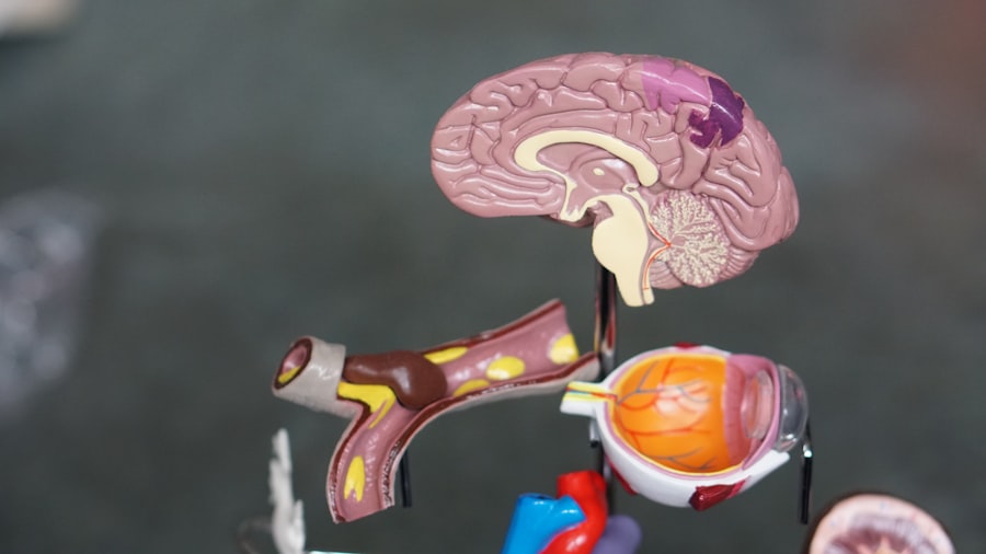
Once differentiation kicks in, the sheer diversity of cell types within your body becomes apparent. Each organ is a complex assembly of many different kinds of cells, each with a unique structure and function, all working in concert.
The Diversity of Cellular Architecture
Consider the liver, a marvel of cellular engineering. It’s not just made of one type of cell. You have hepatocytes, the primary functional cells responsible for metabolism, detoxification, and producing bile. Then there are Kupffer cells, the resident macrophages, acting as the organ’s immune surveillance system. Endothelial cells line the blood vessels, facilitating nutrient and waste exchange, while stellate cells play a role in vitamin storage and fibrosis. This cellular diversity is not accidental; it’s a fundamental requirement for the organ’s multifaceted job.
Beyond Just Structure: Functionality is Key
The unique shape and internal organization of each cell type directly contribute to its function. For instance, the long, slender shape of neurons allows them to transmit electrical signals across vast distances in your nervous system. The flattened shape of red blood cells maximizes their surface area for oxygen transport. The tightly packed structure of epithelial cells in your skin forms a protective barrier. Every cellular adaptation is a testament to evolutionary innovation and the demands of its specific role.
Intercellular Communication: The Essential Dialogue
Cells don’t operate in isolation. They are constantly communicating with each other, sending and receiving signals that coordinate their activities. This intercellular communication is absolutely vital for organ construction. Cells release signaling molecules (like hormones and growth factors) that act like messages, influencing the behavior of neighboring cells. They also form direct connections, allowing for rapid and precise information transfer. Imagine each construction site foreman constantly on the phone, coordinating with other teams.
The Framework: Extracellular Matrix and Scaffolding
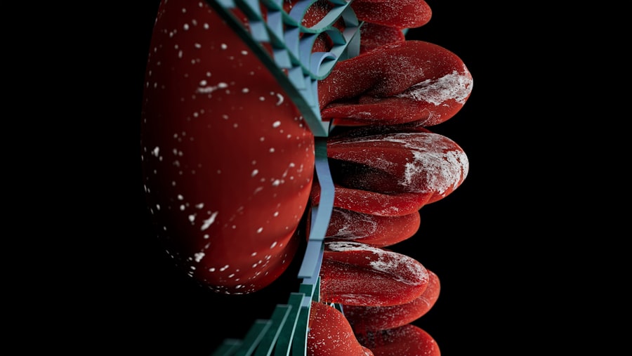
While cells are the active builders, they don’t build organs in a vacuum. They are supported and guided by a complex network of molecules and fibers known as the extracellular matrix (ECM). Think of the ECM as the scaffolding, the reinforcement, and even the mortar that holds the cellular “bricks” together.
The Composition of the ECM: More Than Just Glue
The ECM is not a uniform substance. It’s a dynamic and intricate network composed of various proteins, carbohydrates, and other molecules. Key components include collagen, which provides tensile strength; elastin, which allows tissues to stretch and recoil; and proteoglycans, which help to bind water and create a gel-like environment. The specific composition of the ECM varies greatly depending on the organ and its function, reflecting the diverse mechanical and biological needs of different tissues.
ECM as a Structural Support System
The ECM provides essential structural integrity to organs, preventing them from collapsing and enabling them to withstand mechanical forces. For example, the collagen-rich ECM in your tendons provides the strength needed to connect muscles to bones and transmit forces. In your bones, the ECM, hardened with minerals, creates a rigid framework that supports your entire body. This structural support is crucial even during the delicate stages of organ development.
ECM as a Signaling Hub
The ECM is not just inert scaffolding; it’s an active participant in cellular processes. It can bind to growth factors and other signaling molecules, concentrating them and presenting them to cells, thereby influencing cell behavior, growth, and differentiation. Cells also have receptors that bind to ECM components, transmitting signals into the cell that regulate gene expression and cellular functions. This intricate interplay between cells and their ECM environment is fundamental to coordinated organ development.
Understanding how cells build organs is a fascinating area of study that reveals the intricate processes of development and organization within living organisms. For those interested in exploring this topic further, a related article can provide valuable insights into the mechanisms that guide cellular differentiation and tissue formation. You can read more about these processes in the article found here, which delves into the complexities of how cells work together to create the diverse structures that make up our bodies.
The Assembly Line: Tissue Formation and Organogenesis
| Organ | Cell Types Involved | Key Processes |
|---|---|---|
| Heart | Cardiomyocytes, endothelial cells | Cell proliferation, differentiation, and migration |
| Liver | Hepatocytes, Kupffer cells | Regeneration, metabolic function |
| Brain | Neurons, glial cells | Neurogenesis, synaptogenesis |
| Lungs | Alveolar epithelial cells, endothelial cells | Alveolar development, gas exchange |
With the blueprint, the specialized building blocks, and the scaffolding in place, the remarkable process of organogenesis – the formation of organs – begins in earnest. This involves the coordinated organization of cells and matrix into functional tissues, which then arrange themselves into the complex architecture of organs.
From Cells to Tissues: The First Level of Organization
Tissues are groups of similar cells that work together to perform a specific function. For instance, muscle tissue is composed of muscle cells, while nervous tissue is made up of neurons and glial cells. The formation of these distinct tissues is a crucial intermediate step in organ development, allowing for specialization and the pooling of cellular resources for particular tasks.
The Folding and Shaping of Organs
As tissues mature, they begin to fold, invaginate, and organize into the characteristic shapes of organs. This involves precise patterns of cell growth, migration, and programmed cell death (apoptosis), which sculpts the organ into its final form. Imagine a sheet of paper being folded and cut to create a complex origami figure. This intricate shaping is guided by genetic programs and physical cues.
Vascularization: Supplying the Lifeline
No organ can function without a blood supply. A critical part of organogenesis is the development of blood vessels – a process called vascularization. New blood vessels sprout from existing ones, branching out to provide oxygen and nutrients to the developing tissue and removing waste products. This intricate network of vessels ensures that every cell within the organ receives the essential resources for survival and function.
Innervation: Connecting to the Nervous System
Similarly, organs need to be connected to your nervous system to receive signals and coordinate their actions. Nerves extend into developing organs, establishing communication pathways that allow for control and feedback. This innervation is essential for regulating organ function, from the beating of your heart to the churning of your stomach.
In essence, cellular construction is a breathtaking symphony of coordinated events, orchestrated by your genes and executed by your cells. From the initial genetic blueprint to the intricate assembly of specialized tissues, every step is a testament to the remarkable power of biological engineering. You are not simply a collection of organs; you are a living testament to the continuous, intricate, and awe-inspiring process of cellular construction.

Your Body Knows Something You Don’t
FAQs
What is the process by which cells build organs?
Cells build organs through a process called organogenesis, which involves the coordinated growth, differentiation, and organization of cells to form functional organs. This process is tightly regulated by genetic and environmental factors.
What are the key cellular processes involved in organ development?
Key cellular processes involved in organ development include cell proliferation, cell differentiation, cell migration, and cell signaling. These processes work together to create the complex structures and functions of organs.
How do cells communicate and coordinate during organ development?
Cells communicate and coordinate during organ development through various signaling pathways, including chemical signals such as growth factors, hormones, and neurotransmitters. These signals help cells to make decisions about their fate, position, and function within the developing organ.
What role do stem cells play in organ development?
Stem cells play a crucial role in organ development by giving rise to the different cell types that make up the organ. They have the ability to self-renew and differentiate into specialized cell types, contributing to the growth and repair of organs throughout development and adulthood.
How do genetic and environmental factors influence organ development?
Genetic factors, such as the expression of specific genes, and environmental factors, such as nutrient availability and exposure to toxins, can influence organ development. These factors can impact cell behavior, differentiation, and overall organ structure and function.