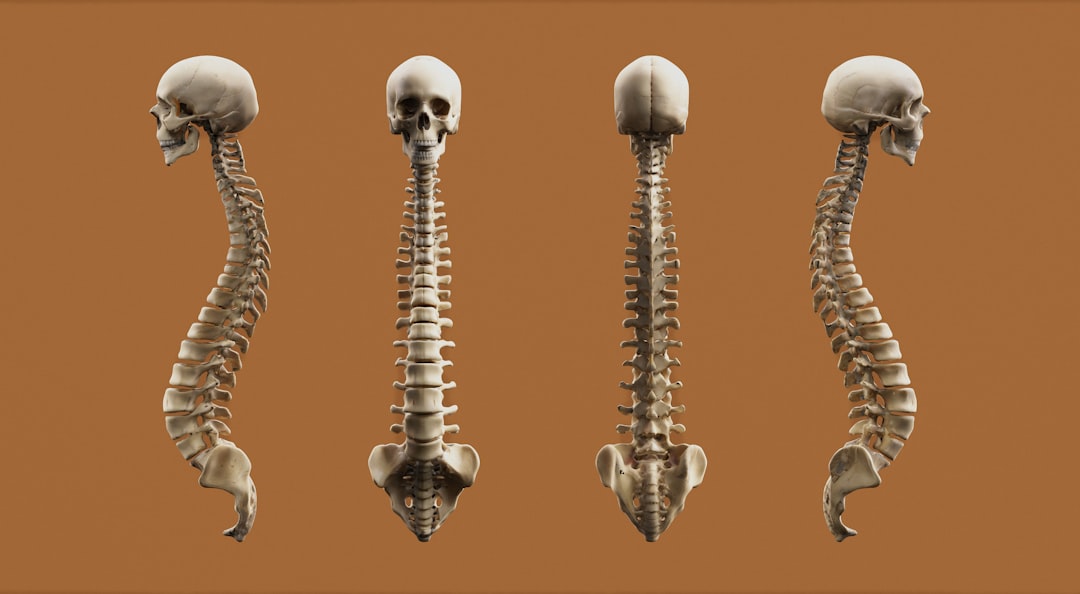
You might not think of your immune system when you consider bone health, but it plays a surprisingly significant role. Specifically, a specialized type of immune cell, the regulatory T (Treg) cell, and the markers it expresses are key players in the intricate process of bone formation and maintenance. Understanding how these elements interact can shed light on conditions where bone becomes compromised and points toward potential therapeutic avenues.
Your immune system, often perceived as a defense against pathogens, also acts as a crucial regulator of tissue. It’s not just about attacking invaders; it’s also about maintaining balance and ensuring that your body’s own tissues function correctly and are repaired when necessary. This is where Treg cells come into play, acting as the immune system’s peacekeepers and facilitators of the delicate equilibrium, or homeostasis, that governs your tissues, including bone.
Defining Treg Cells: More Than Just Immuno-Suppression
Treg cells are a distinct subset of T lymphocytes, a type of white blood cell. Their primary function is to dampen immune responses, preventing the immune system from overreacting and causing damage to the body’s own tissues. While this immunosuppressive role is central, it’s not the complete picture. Treg cells are increasingly recognized for their broader involvement in tissue repair and regeneration. They don’t simply shut down the immune system; they modulate its activity to create an environment conducive to healing and proper function.
Identifying Treg Cells: The Foxp3 Hallmark
A defining characteristic of most Treg cells is the expression of a transcription factor called Forkhead box P3 (Foxp3). This molecule is indispensable for the development and function of these cells. You can consider Foxp3 as the master switch that dictates the Treg cell lineage. Without it, T cells targeted to become Tregs would differentiate into other types of immune cells. Therefore, the presence of Foxp3 is a primary reliable indicator for identifying Treg cells in various tissues, including the bone microenvironment.
Other Surface Markers: A Supporting Cast of Characters
While Foxp3 is intracellular, other markers are found on the surface of Treg cells, making them easier to detect and study. These surface markers are often used in conjunction with Foxp3 to further characterize Treg populations.
CD4 and CD25: The Ubiquitous Duo
Most Treg cells are CD4+ T cells, meaning they express the CD4 co-receptor on their surface. CD4 is also found on helper T cells, so this marker alone isn’t sufficient to identify Tregs. However, a significant majority of Treg cells also express high levels of CD25, the alpha chain of the interleukin-2 receptor. CD25 is crucial for Treg cell survival and function, as it allows them to bind to interleukin-2 (IL-2), a potent immune-stimulating cytokine. The combination of CD4 and high CD25 expression is a common, though not exclusive, way to identify potential Treg cells.
Helios and Neuropilin-1: Nuances in Identification
Further refinement in identifying Treg cells involves markers like Helios and Neuropilin-1 (Nrp-1). Helios is a transcription factor that is often co-expressed with Foxp3 in thymic Treg cells. Its presence can help distinguish between naturally arising Tregs and those induced in the periphery. Nrp-1, a transmembrane receptor, has also been implicated in Treg cell function and suppressive capacity, particularly in certain tissue environments. Identifying these markers alongside Foxp3 and CD25 provides a more detailed understanding of the specific Treg populations present.
Recent studies have highlighted the intriguing relationship between regulatory T cells (Treg cells) and bone formation markers, suggesting that Treg cells may play a crucial role in bone metabolism and health. For a deeper understanding of this connection, you can explore the article available at Freaky Science, which discusses the implications of Treg cells in the regulation of osteoblast activity and their potential impact on bone density and overall skeletal health.
Bone Remodeling: A Constant Dance Between Formation and Resorption
Your skeleton is not a static structure; it’s a dynamic tissue undergoing constant remodeling throughout your life. This process involves the coordinated action of cells that build bone and cells that break it down. Maintaining the right balance between these two processes is paramount for strong, healthy bones. When this balance is disrupted, it can lead to a range of skeletal disorders.
Osteoblasts: The Bone Builders
Osteoblasts are the specialized cells responsible for synthesizing and depositing new bone matrix, primarily composed of collagen and other proteins. This process is known as osteogenesis. These cells are the architects of your skeleton, laying down the foundational material for bone.
Differentiation from Mesenchymal Stem Cells
Osteoblasts originate from mesenchymal stem cells (MSCs), multipotent cells residing in various tissues, including the bone marrow. Under specific signaling cues, these MSCs differentiate into osteoprogenitor cells, which then mature into fully functional osteoblasts. The journey from a pluripotent stem cell to a bone-forming cell is a complex cascade of gene expression and cellular events.
Key Markers of Osteoblast Activity
You can identify osteoblasts and assess their activity by looking for specific markers they express. These markers serve as indicators of their developmental stage and their functional state.
Alkaline Phosphatase (ALP): A Metabolic Indicator
Alkaline phosphatase is an enzyme produced by osteoblasts. Its activity is closely linked to the process of bone mineralization, where calcium and phosphate are deposited into the bone matrix. Elevated levels of ALP in the blood or localized ALP activity in bone tissue are often indicative of active bone formation.
Osteocalcin: The Bone Matrix Protein
Osteocalcin is a non-collagenous protein secreted by mature osteoblasts. It plays a critical role in bone mineralization and the regulation of bone turnover. Osteocalcin is a useful marker for assessing the rate of bone formation in both research settings and clinical diagnostics.
Type I Collagen (Col I): The Structural Backbone
Type I collagen is the most abundant protein in bone and forms the structural framework of the bone matrix. Osteoblasts are responsible for its synthesis and secretion. While Col I is a general marker of connective tissue, its specific organization and deposition within the bone matrix are unique to osteoblast activity.
Osteoclasts: The Bone Resorbers
In contrast to osteoblasts, osteoclasts are multinucleated cells responsible for bone resorption, the process of breaking down and removing old or damaged bone tissue. This is a crucial step in the remodeling cycle, clearing space for new bone to be formed.
Origin from Hematopoietic Precursors
Unlike osteoblasts, osteoclasts arise from hematopoietic stem cells, the same lineage that gives rise to blood cells. These precursors fuse together to form the large, multinucleated osteoclasts. Their development and activation are tightly regulated by signaling molecules within the bone microenvironment.
Key Markers of Osteoclast Activity
Similar to osteoblasts, specific markers can identify osteoclasts and their functional status.
Tartrate-Resistant Acid Phosphatase (TRAP): A Signature Enzyme
Tartrate-resistant acid phosphatase (TRAP) is an enzyme characteristically found in osteoclasts. Its presence is a widely accepted marker for identifying these bone-resorbing cells. TRAP is often used in histological studies to visualize and quantify osteoclast populations.
Cathepsin K: The Collagenase Enzyme
Cathepsin K is a potent cysteine protease that plays a significant role in the degradation of the bone matrix, particularly type I collagen. It is highly expressed in mature osteoclasts and is essential for their bone-resorbing capabilities. Inhibition of cathepsin K is a target for treating osteoporosis.
Carbonic Anhydrase II: The Proton Pump Component
Carbonic anhydrase II is an enzyme involved in the production of protons (H+) by osteoclasts. These protons are secreted into the resorption lacuna, creating an acidic environment that decalcifies the bone matrix, making it susceptible to enzymatic digestion.
Treg Cells and Their Influence on Bone Cells

The presence and activity of Treg cells within the bone marrow and surrounding bone tissue directly impact the behavior of osteoblasts and osteoclasts. This interaction is not merely coincidental; it’s a critical mechanism for maintaining bone homeostasis. You can think of Treg cells as modulators, fine-tuning the bone remodeling process.
Direct Interaction with Osteoblasts
Treg cells can directly interact with osteoblasts, influencing their differentiation, proliferation, and function. These interactions can lead to either promotion or suppression of bone formation, depending on the context and the specific signals exchanged.
Modulation of Osteoblast Differentiation and Proliferation
Treg cells can secrete cytokines and other signaling molecules that either encourage or inhibit the differentiation of osteoprogenitor cells into mature osteoblasts. They can also influence how many osteoblasts are produced and how long they survive. For instance, some studies suggest that certain Treg cell subsets can promote osteoblastogenesis, contributing to bone formation.
Inhibition of Osteoblast Apoptosis
In some situations, Treg cells might protect osteoblasts from programmed cell death (apoptosis). By preventing the premature demise of bone-building cells, they can help maintain a pool of active osteoblasts, thereby supporting bone formation.
Regulation of Osteoclastogenesis and Activity
Perhaps the most well-established role of Treg cells in bone is their influence over osteoclasts. By suppressing osteoclast formation and activity, Treg cells prevent excessive bone breakdown.
Suppressing Osteoclast Differentiation and Survival
Treg cells can significantly reduce the number of osteoclasts that develop from their precursors. They achieve this by interfering with signaling pathways essential for osteoclast differentiation and by promoting osteoclast apoptosis. This direct suppression of osteoclast development is a key mechanism by which Tregs protect bone from excessive resorption.
Inhibiting Osteoclast Function
Even once osteoclasts are formed, Treg cells can dampen their bone-resorbing activity. This can involve interfering with the signals that activate osteoclasts or directly inhibiting their ability to attach to the bone surface and secrete enzymes.
Key Markers of Treg Cell Activity in the Bone Microenvironment
To understand how Treg cells influence bone, researchers look for specific markers that indicate their presence and activity within the bone microenvironment. These markers provide valuable insights into the role of Tregs in various bone conditions.
Foxp3 as a Primary Indicator
As mentioned earlier, Foxp3 is the linchpin for Treg cell identity. Within the bone microenvironment, the presence of Foxp3+ cells is a strong indicator of Treg cell infiltration and potential influence.
Localization within Bone Marrow and Trabecular Bone
You can find Foxp3+ cells within the bone marrow cavity, interspersed among hematopoietic stem cells and stromal cells. They can also be detected in the vicinity of the trabecular bone surface, meaning they are physically close to where bone remodeling is actively occurring, allowing for direct interactions with osteoblasts and osteoclasts.
Correlation with Bone Density
Studies have shown correlations between the number of Foxp3+ cells in bone tissue and bone density. In general, a higher presence of these cells is often associated with better bone health, suggesting their protective role. Conversely, a decrease in Foxp3+ cells can be linked to conditions characterized by bone loss.
Other Treg-Associated Markers in Bone
Beyond Foxp3, other markers help delineate Treg cell function and their specific subtypes within the bone microenvironment.
CTLA-4: A Key Inhibitory Receptor
Cytotoxic T-lymphocyte-associated protein 4 (CTLA-4) is a crucial inhibitory receptor expressed on Treg cells. It plays a significant role in dampening immune responses by competing with the co-stimulatory receptor CD28 for binding to ligands on antigen-presenting cells. High expression of CTLA-4 on bone-associated Treg cells is often indicative of their suppressive capacity and their contribution to maintaining immune tolerance within the bone tissue.
IL-10: The Immunomodulatory Cytokine
Interleukin-10 (IL-10) is a pleiotropic cytokine known for its potent immunosuppressive and anti-inflammatory properties. Treg cells are a major source of IL-10, and this cytokine plays a vital role in regulating bone remodeling. IL-10 can inhibit osteoclast differentiation and activity while promoting osteoblast function. Therefore, detecting IL-10 within the bone microenvironment, particularly in association with Treg cells, signifies their active modulation of bone homeostasis.
TGF-β: A Multifaceted Regulator
Transforming growth factor-beta (TGF-β) is another critical cytokine involved in various cellular processes, including bone metabolism. Treg cells can produce and respond to TGF-β, and it can influence both osteoblast and osteoclast function. While its effects are complex and can be context-dependent, TGF-β is frequently implicated in the interplay between the immune system and bone, with Treg cells acting as key mediators.
Recent studies have highlighted the intriguing relationship between T regulatory (Treg) cells and bone formation markers, suggesting that these immune cells may play a significant role in bone health and regeneration. For a deeper understanding of this connection, you can explore a related article that discusses the impact of Treg cells on osteoblast activity and their potential therapeutic implications. This research opens new avenues for treating bone-related disorders and emphasizes the importance of immune regulation in skeletal biology. To read more about this fascinating topic, visit this article.
Clinical Significance: Treg Cells and Bone Pathologies
| Marker | Treg Cells | Bone Formation |
|---|---|---|
| CD25 | Increased expression | Stimulates osteoblast differentiation |
| FOXP3 | Regulates Treg cell function | Enhances bone formation |
| RANKL | Suppresses RANKL expression | Regulates osteoclast activity |
The intricate relationship between Treg cells and bone remodeling has significant implications for understanding and treating a range of bone pathologies. When this delicate balance is disrupted, it can contribute to the development or exacerbation of diseases characterized by bone loss or abnormal bone formation.
Osteoporosis: When Bone Resorption Outpaces Formation
Osteoporosis is a systemic skeletal disease characterized by low bone mass and deterioration of bone tissue, leading to increased fragility and risk of fractures. In many cases of osteoporosis, an imbalance exists where bone resorption by osteoclasts exceeds bone formation by osteoblasts.
Treg Cell Deficiency and Osteoporosis Risk
A deficiency in functional Treg cells or impaired Treg cell activity has been linked to an increased risk of developing osteoporosis. When the suppressive influence of Tregs on osteoclasts is diminished, osteoclast activity can become dysregulated, leading to excessive bone breakdown. Conversely, strategies aimed at enhancing Treg cell function or bolstering their numbers have shown promise in preclinical models of osteoporosis.
Therapeutic Potential of Treg Cell Modulation
Targeting Treg cells represents a potential therapeutic strategy for osteoporosis. This could involve:
Enhancing Treg Cell Numbers and Function
Methods to increase the population of functional Treg cells or to boost their inherent suppressive capabilities are being explored. This might involve the use of specific drugs or biological agents that promote Treg cell differentiation and activity.
Utilizing Treg Cell-Derived Molecules
Leveraging the beneficial molecules produced by Treg cells, such as IL-10, could also offer a way to restore bone homeostasis. Administering these cytokines directly or stimulating their endogenous production might help to rebalance bone remodeling.
Rheumatoid Arthritis: Inflammation’s Toll on Bone
Rheumatoid arthritis (RA) is a chronic autoimmune disease characterized by inflammation of the joints, which can lead to significant bone erosion and joint destruction. The inflammatory environment in RA can disrupt the normal balance of bone remodeling.
Treg Cell Dysfunction in RA
In RA, Treg cells often exhibit impaired suppressive function or reduced numbers, contributing to the unchecked immune attack on joint tissues, including bone. This Treg cell dysfunction allows pro-inflammatory cytokines to dominate, promoting osteoclast activation and leading to bone resorption around the affected joints.
Restoring Treg Cell Balance for Joint and Bone Protection
Restoring the balance of Treg cell activity in RA could be crucial for both reducing joint inflammation and preserving bone integrity. Therapies aimed at enhancing Treg cell function could help to dampen the autoimmune response and its detrimental effects on bone.
Other Bone-Related Conditions
Beyond osteoporosis and RA, the role of Treg cells is being investigated in other bone-related conditions, including:
Periodontitis: Gum Disease and Bone Loss
Periodontitis, a severe form of gum disease, involves inflammation and destruction of the supporting structures of the teeth, including the alveolar bone. Dysregulation of Treg cells has been implicated in the progression of periodontitis and the associated bone loss.
Bone Metastases: Cancer’s Impact on Bone
When cancer spreads to the bone (bone metastases), it can significantly disrupt bone homeostasis, leading to pain, fractures, and hypercalcemia. The interactions between cancer cells, the immune system, and bone cells are complex, and Treg cells may play a role in modulating the bone microenvironment in the context of metastasis. Understanding these interactions could identify new therapeutic targets.
In conclusion, you can see that Treg cells, identified by markers like Foxp3, are far more than just immune suppressors. They are integral regulators of bone formation and maintenance. By influencing the delicate interplay between osteoblasts and osteoclasts, and by expressing specific markers that indicate their activity, Treg cells play a critical role in maintaining skeletal health. Research into these cells and their associated markers is vital for developing novel strategies to combat bone diseases.
FAQs
What are Treg cells?
Treg cells, or regulatory T cells, are a type of immune cell that helps maintain immune system balance and prevent autoimmune diseases by suppressing the activity of other immune cells.
How do Treg cells relate to bone formation markers?
Treg cells have been found to play a role in regulating bone metabolism and bone formation. They can influence the activity of osteoclasts and osteoblasts, which are the cells responsible for bone resorption and bone formation, respectively.
What are bone formation markers?
Bone formation markers are substances that are released during the process of bone formation. These markers can be measured in the blood and are used to assess bone health and turnover.
How do Treg cells impact bone formation markers?
Treg cells have been shown to influence the production and activity of bone formation markers. They can modulate the balance between bone resorption and bone formation, ultimately affecting bone density and strength.
What are the implications of the relationship between Treg cells and bone formation markers?
Understanding the role of Treg cells in bone metabolism and their impact on bone formation markers could lead to the development of new treatments for bone-related diseases, such as osteoporosis and rheumatoid arthritis. It also highlights the interconnectedness of the immune system and bone health.